

Secretory immunoglobulin A induces human lung fibroblasts to produce inflammatory cytokines and undergo activation
- PMID: 30570135
- PMCID: PMC6378381
- DOI: 10.1111/cei.13253
Secretory immunoglobulin A induces human lung fibroblasts to produce inflammatory cytokines and undergo activation
Abstract
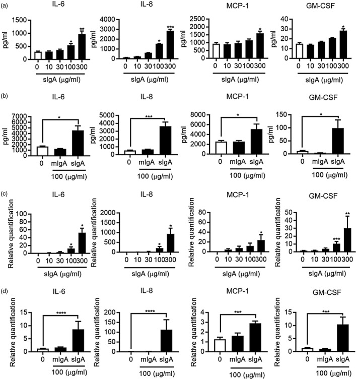
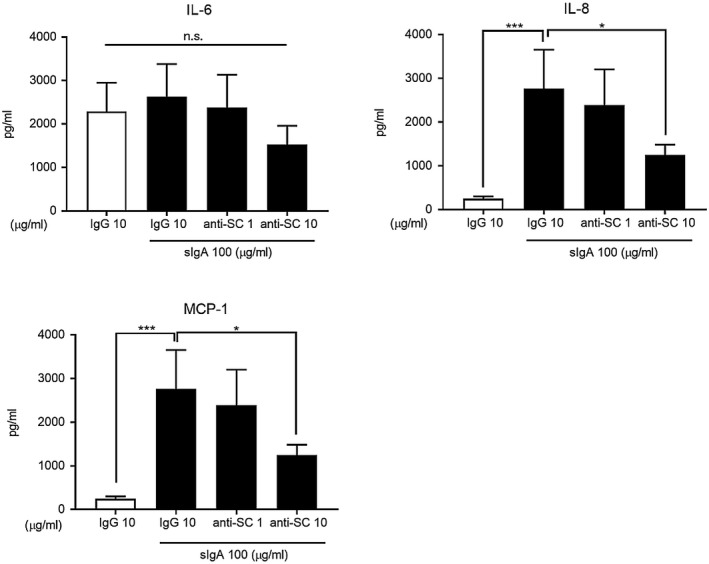
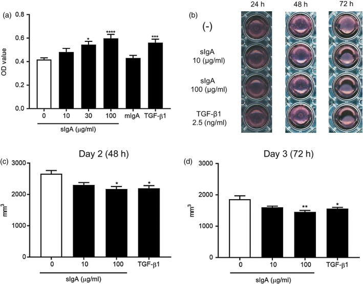
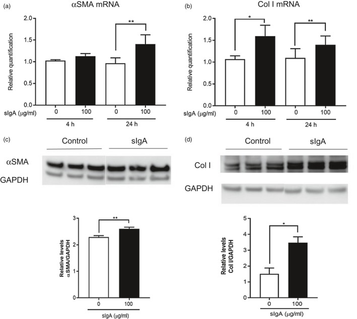
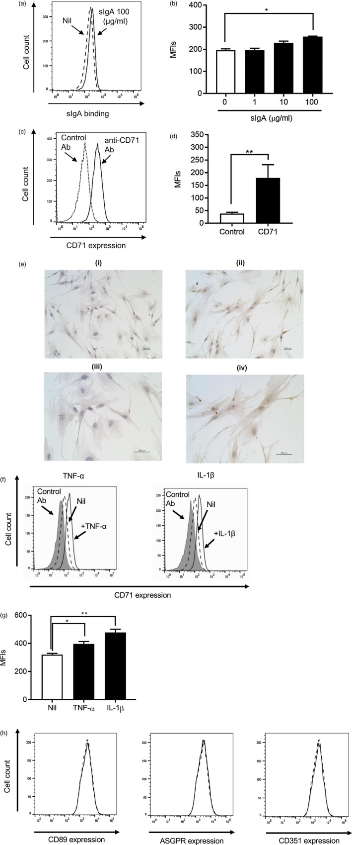
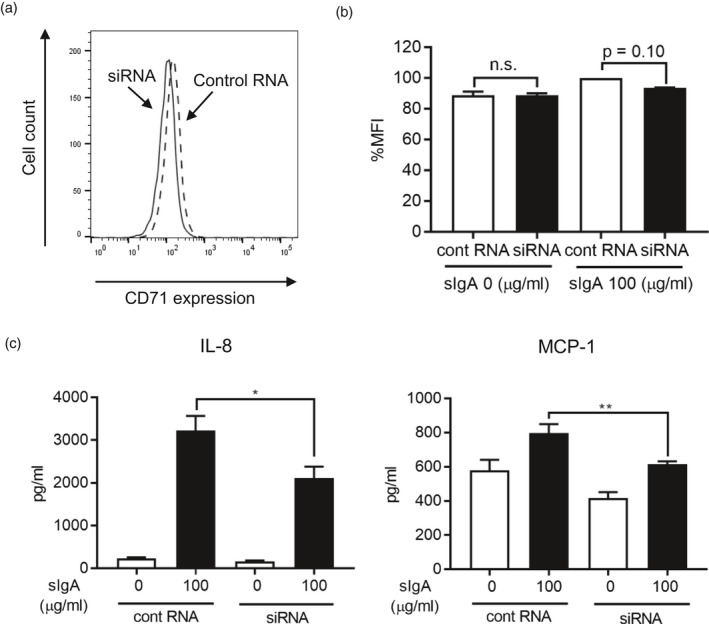
Immunoglobulin (Ig)A is the most abundant immunoglobulin in humans, and in the airway mucosa secretory IgA (sIgA) plays a pivotal role in first-line defense against invading pathogens and antigens. IgA has been reported to also have pathogenic effects, including possible worsening of the prognosis of idiopathic pulmonary fibrosis (IPF). However, the precise effects of IgA on lung fibroblasts remain unclear, and we aimed to elucidate how IgA activates human lung fibroblasts. We found that sIgA, but not monomeric IgA (mIgA), induced interleukin (IL)-6, IL-8, monocyte chemoattractant protein (MCP)-1 and granulocyte-macrophage colony-stimulating factor (GM-CSF) production by normal human lung fibroblasts (NHLFs) at both the protein and mRNA levels. sIgA also promoted proliferation of NHLFs and collagen gel contraction comparable to with transforming growth factor (TGF)-β, which is involved in fibrogenesis in IPF. Also, Western blot analysis and real-time quantitative polymerase chain reaction (PCR) revealed that sIgA enhanced production of α-smooth muscle actin (α-SMA) and collagen type I (Col I) by NHLFs. Flow cytometry showed that NHLFs bound sIgA, and among the known IgA receptors, NHLFs significantly expressed CD71 (transferrin receptor). Transfection of siRNA targeting CD71 partially but significantly suppressed cytokine production by NHLFs co-cultured with sIgA. Our findings suggest that sIgA may promote human lung inflammation and fibrosis by enhancing production of inflammatory or fibrogenic cytokines as well as extracellular matrix, inducing fibroblast differentiation into myofibroblasts and promoting human lung fibroblast proliferation. sIgA's enhancement of cytokine production may be due partially to its binding to CD71 or the secretory component.
Keywords: IgA; fibroblasts; fibrosis; human; lung.
© 2018 British Society for Immunology.
Figures
Similar articles
-
Secretory IgA accumulated in the airspaces of idiopathic pulmonary fibrosis and promoted VEGF, TGF-β and IL-8 production by A549 cells.Clin Exp Immunol. 2020 Mar;199(3):326-336. doi: 10.1111/cei.13390. Epub 2019 Nov 8. Clin Exp Immunol. 2020. PMID: 31660581 Free PMC article.
-
Leptin enhances cytokine/chemokine production by normal lung fibroblasts by binding to leptin receptor.Allergol Int. 2019 Sep;68S:S3-S8. doi: 10.1016/j.alit.2019.04.002. Epub 2019 Apr 24. Allergol Int. 2019. PMID: 31029506
-
Proliferation and Cytokine Production of Human Mesangial Cells Stimulated by Secretory IgA Isolated from Patients with IgA Nephropathy.Cell Physiol Biochem. 2015;36(5):1793-1808. doi: 10.1159/000430151. Cell Physiol Biochem. 2015. PMID: 26184511
-
Could the airway epithelium play an important role in mucosal immunoglobulin A production?Clin Exp Allergy. 1999 Dec;29(12):1597-605. doi: 10.1046/j.1365-2222.1999.00644.x. Clin Exp Allergy. 1999. PMID: 10594535 Review.
-
SIgA in various pulmonary diseases.Eur J Med Res. 2023 Aug 27;28(1):299. doi: 10.1186/s40001-023-01282-5. Eur J Med Res. 2023. PMID: 37635240 Free PMC article. Review.
Cited by
-
Distinct features of SARS-CoV-2-specific IgA response in COVID-19 patients.Eur Respir J. 2020 Aug 27;56(2):2001526. doi: 10.1183/13993003.01526-2020. Print 2020 Aug. Eur Respir J. 2020. PMID: 32398307 Free PMC article.
-
Humoral immune mechanisms involved in protective and pathological immunity during COVID-19.Hum Immunol. 2021 Oct;82(10):733-745. doi: 10.1016/j.humimm.2021.06.011. Epub 2021 Jul 1. Hum Immunol. 2021. PMID: 34229864 Free PMC article. Review.
-
Enhanced Bruton's tyrosine kinase in B-cells and autoreactive IgA in patients with idiopathic pulmonary fibrosis.Respir Res. 2019 Oct 24;20(1):232. doi: 10.1186/s12931-019-1195-7. Respir Res. 2019. PMID: 31651327 Free PMC article.
-
Serum levels of the IgA isotype switch factor TGF-β1 are elevated in patients with COVID-19.FEBS Lett. 2021 Jul;595(13):1819-1824. doi: 10.1002/1873-3468.14104. Epub 2021 May 21. FEBS Lett. 2021. PMID: 33961290 Free PMC article.
-
Anti-SARS-CoV-2 Immunoglobulin Isotypes, and Neutralization Activity Against Viral Variants, According to BNT162b2-Vaccination and Infection History.Front Immunol. 2021 Dec 17;12:793191. doi: 10.3389/fimmu.2021.793191. eCollection 2021. Front Immunol. 2021. PMID: 34975897 Free PMC article.
References
-
- Mestecky J, Russell MW, Jackson S, Brown TA. The human IgA system: A reassessment. Clin Immunol Immunopathol 1986; 40:105–14. - PubMed
-
- Newkirk MM, Klein MH, Katz A, Fisher MM, Underdown BJ. Estimation of polymeric IgA in human serum: An assay based on binding of radiolabeled human secretory component with applications in the study of IgA nephropathy, IgA monoclonal gammopathy, and liver disease. J Immunol (Balt) 1983; 130:1176–81. - PubMed
-
- Stockley RA, Afford SC, Burnett D. Assessment of 7S and 11S immunoglobulin A in sputum. Am Rev Respir Dis 1980; 122:959–64. - PubMed
-
- Puchelle E, Jacqot J, Zahm JM. In vitro restructuring effect of human airway immunoglobulins A and lysozyme on airway secretions. Eur J Respir Dis Suppl 1987; 153:117–22. - PubMed
Publication types
MeSH terms
Substances
LinkOut - more resources
Full Text Sources
Miscellaneous
