. 2019 Apr;137(4):585-598.
doi: 10.1007/s00401-018-1947-3. Epub 2018 Dec 20.

Seeding selectivity and ultrasensitive detection of tau aggregate conformers of Alzheimer disease

Affiliations

Seeding selectivity and ultrasensitive detection of tau aggregate conformers of Alzheimer disease

Allison Kraus et al. Acta Neuropathol. 2019 Apr.

Abstract

Alzheimer disease (AD) and chronic traumatic encephalopathy (CTE) involve the abnormal accumulation in the brain of filaments composed of both three-repeat (3R) and four-repeat (4R) (3R/4R) tau isoforms. To probe the molecular basis for AD's tau filament propagation and to improve detection of tau aggregates as potential biomarkers, we have exploited the seeded polymerization growth mechanism of tau filaments to develop a highly selective and ultrasensitive cell-free tau seed amplification assay optimized for AD (AD real-time quaking-induced conversion or AD RT-QuIC). The reaction is based on the ability of AD tau aggregates to seed the formation of amyloid fibrils made of certain recombinant tau fragments. AD RT-QuIC detected seeding activity in AD (n = 16) brains at dilutions as extreme as 107-1010-fold, but was 102-106-fold less responsive when seeded with brain from most cases of other types of tauopathy with comparable loads of predominant 3R or 4R tau aggregates. For example, AD brains had average seeding activities that were orders of magnitude higher than Pick disease brains with predominant 3R tau deposits, but the opposite was true using our previously described Pick-optimized tau RT-QuIC assay. CTE brains (n = 2) had seed concentrations comparable to the weakest of the AD specimens, and higher than 3 of 4 specimens with 3R/4R primary age-related tauopathy. AD seeds shared properties with the tau filaments found in AD brains, as AD seeds were sarkosyl-insoluble, protease resistant, and reactive with tau antibodies. Moreover, AD RT-QuIC detected as little as 16 fg of pure synthetic tau fibrils. The distinctive seeding activity exhibited by AD and CTE tau filaments compared to other types of tauopathies in these seeded polymerization reactions provides a mechanistic basis for their consistent propagation as specific conformers in patients with 3R/4R tau diseases. Importantly, AD RT-QuIC also provides rapid ultrasensitive quantitation of 3R/4R tau-seeding activity as a biomarker.

Keywords: Alzheimer disease; Biomarker; Chronic traumatic encephalopathy; Diagnosis; RT-QuIC; Seeds; Tau aggregate; Tauopathy.


Conflict of interest statement

Conflict of interest

AK, ES, MM, and BC are named inventors on a PCT patent application (PCT/US2017/069024) related to the technology described herein. The other authors declare that they have no other competing interests.

Ethics approval

None required; all samples analyzed were from deceased, de-identified individuals. Nonetheless, exemption #13437 from Office of Human Subjects Research was obtained by BC.

Consent for publication

Not applicable

Availability of data and material

All data generated or analyzed during this study are included in this published article [and its supplementary information files].

Figures
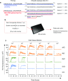
Fig. 1
Fig. 1
AD RT-QuIC dilution analyses of AD (3R/4R tauopathy), PSP (4R tauopathy), CVD (histologically negative for tau pathology), and KO (tau-free) brain homogenates. a Schematic of τ306 and K19CFh with His-tags and the S322C mutation denoted, with numbering based on the full-length htau40 sequence. b Reactions were seeded with 1 µL of brain homogenate at the indicated dilution in a 384-well plate, subjected to cycles of shaking and rest, and periodically measured for relative ThT fluorescence over 30 h. c Each curve represents an individual well, run in quadruplicate for each dilution
Fig. 2
Fig. 2
Lag time analysis of AD RT-QuIC reactions seeded with dilutions of AD and non-AD brain homogenates. Lag time was determined as the reaction time required to exceed a ThT fluorescence threshold of the average + 100 standard deviations of the baseline fluorescence. Symbols indicate lag times from individual wells. Cross hatches and bars indicate the mean ± SD of the values at each dilution. The assay endpoint was 30 h, and thus, any data points beyond the red line had positive ThT fluorescence values at or greater than 30 h. A value of 30 was assigned to data points beyond the assay endpoint to calculate mean ± SD
Fig. 3
Fig. 3
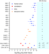
AD RT-QuIC end-point dilution analysis of brain homogenates from cases of AD, CTE, and other neurological disorders. The seeding dose (SD50) was determined by Spearman–Kärber analyses and is shown as log SD50/mg brain tissue. The vertical grey and blue lines mark the average values from brains of tau knock-out (KO) mice and human CVD cases lacking immunohistochemical evidence of tau pathology, respectively. sAD sporadic AD, fAD familial AD, CTE chronic traumatic encephalopathy, PiD Pick disease, PSP progressive supranuclear palsy, CBD corticobasal degeneration, AGD argyrophilic grain disease, FTDP-17 frontotemporal dementia and Parkinsonism-17, SC senile changes (non-tau associated), CVD cerebrovascular disease, DLBD diffuse Lewy body disease, FTLD-TDP frontotemporal lobar degeneration with TDP-43, ALS amyotrophic lateral sclerosis, PD Parkinson disease, IHC immunohistochemistry. Data are represented as mean ± SD. sAD, fAD, p < 0.0001; CTE, p < 0.0001; PART, p=0.0002; CBD, p = 0.005; AGD, p=0.04, PiD, p=0.01; DLBD, p=0.015 by one-way ANOVA [F(15, 31) = 29.15] compared to CVD
Fig. 4
Fig. 4
Tau antibody reactivity, detergent insolubility, and protease resistance of AD seeding activity. a Serial immunodepletion. HT7 anti-tau and control IgG antibody-conjugated beads were incubated with sAD brain homogenate and two rounds of unbound (supernatant) fractions were assayed by end-point dilution in the AD RT-QuIC. b Immune capture. Bound (then eluted from beads) and unbound (supernatant) fractions of sAD brain homogenate were assayed by end-point dilution in the AD RT-QuIC. Mean log SD50 values (± SE) were calculated using Spearman–Kärber analysis. c Dilution analysis of total and sarkosyl-insoluble fractions derived from identical brain equivalents of CVD, DLBD, and AD brain homogenates. Lag times from individual reactions seeded with the designated dilutions are shown. Horizontal bars indicate the mean ± SD of quadruplicate lag times. d AD, DLBD, and CVD brain homogenates were digested with proteinase K (+PK) and used for dilution analysis. Lag times are shown for untreated (total) and proteinase K-treated brain homogenates. For c, d, any data points beyond the assay endpoint (indicated by a red line) had positive ThT fluorescence values at or greater than 30 h. These data points were assigned a value of 30 to calculate mean ± SD
Fig. 5
Fig. 5
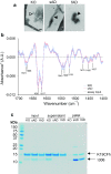
AD RT-QuIC products are fibrillar, high in β-sheet and insoluble. a Negative stain electron micrographs of products of AD RT-QuIC reactions seeded with KO, sAD or fAD brain homogenates. Open arrow head: amorphous aggregates in KO-seeded reactions; Closed arrowheads: fibrils in AD-seeded reactions. Asterisks: EM grid structure. b Average ± SD of second derivative FTIR spectra of proteinase K-treated RT-QuIC substrate input and recovered products are shown. RT-QuIC products used for analysis were initially seeded with fAD or sAD as indicated. Prominent bands at 1630 and 1617 cm−1 are consistent with β-sheet secondary structure. c RT-QuIC products were collected after 20 h of incubation, pelleted, and compared to the starting reaction (input) and supernatant fractions using gel analysis. Pellets were concentrated ~ fivefold compared to the total reaction. K19CFh and τ306 are denoted with arrows
Fig. 6
Fig. 6
AD RT-QuIC dilution analyses of cortex tissue homogenates from brain samples with different Braak stages. a End-point dilution analyses were carried out on brain tissue homogenates from the cortex of sAD (Braak stage V or VI) and non-demented but with Braak stage II or III pathology samples. Results are shown as log SD50/mg brain tissue. b Lag time, determined by the assay time required to exceed ThT fluorescence values of the average + 100 standard deviations of the baseline fluorescence. Each symbol represents an individual well. Cross hatches and bars indicate the mean ± SD of the values at each dilution. Any data points beyond the assay endpoint (indicated by a red line) had positive ThT fluorescence values at or greater than 30 h. These data points were assigned a value of 30 to calculate mean ± SD
Fig. 7
Fig. 7
Seeding activity in brain regions with and without immunohistochemically visible 3R/4R tau deposits. Seeding activity measured in brain tissue homogenates derived from the frontal cortex, precuneus/posterior cingulate (PPC) cortex, temporal cortex and cerebellum from AD, CTE, and PART cases is indicated. Results are shown as log SD50/mg brain tissue (mean ± SD; n = 1–5 independent end-point dilution analyses for each case) and are originally plotted in Fig. 3 for all cases except select brain regions from PART 4 and AD 9 and 10 cases. One PART and two AD cases had neuropathological diagnoses of cerebral amyloid angiopathy (CAA) in the cerebellum and frontal cortex. Seeding activity was measured in homogenates from both the cerebellum, which lacks immunochemically visible tau deposits, and the frontal cortex. For the PART case, seeding activity was also determined for a homogenate derived from the temporal cortex. AD Alzheimer disease, CTE chronic traumatic encephalopathy, PART primary age-related tauopathy, PPC precuneus/posterior cingulate cortex, C cerebellum, F frontal cortex, T temporal cortex, IHC immunohistochemistry, SD50 seeding dose

References

    1. Atarashi R, Satoh K, Sano K, Fuse T, Yamaguchi N, Ishibashi D, et al. Ultrasensitive human prion detection in cerebrospinal fluid by real-time quaking-induced conversion. Nat Med. 2011;17:175–178. doi: 10.1038/nm.2294. - DOI - PubMed
    1. Bongianni M, Orrù CD, Groveman BR, Sacchetto L, Fiorini M, Tonoli G, et al. Diagnosis of human prion disease using real-time quaking-induced conversion testing of olfactory mucosa and cerebrospinal fluid samples. JAMA Neurol. 2017;74:1–8. doi: 10.1001/jamaneurol.2016.4614. - DOI - PubMed
    1. Braak H, Del Tredici K, Rub U, de Vos RA, Jansen Steur EN, Braak E. Staging of brain pathology related to sporadic Parkinson’s disease. Neurobiol Aging. 2003;24:197–211. doi: 10.1016/S0197-4580(02)00065-9. - DOI - PubMed
    1. Cairns NJ, Bigio EH, Mackenzie IR, Neumann M, Lee VM, Hatanpaa KJ, et al. Neuropathologic diagnostic and nosologic criteria for frontotemporal lobar degeneration: consensus of the Consortium for Frontotemporal Lobar Degeneration. Acta Neuropathol. 2007;114:5–22. doi: 10.1007/s00401-007-0237-2. - DOI - PMC - PubMed
    1. Castilla J, Saa P, Morales R, Abid K, Maundrell K, Soto C. Protein misfolding cyclic amplification for diagnosis and prion propagation studies. Methods Enzymol. 2006;412:3–21. doi: 10.1016/S0076-6879(06)12001-7. - DOI - PubMed

Publication types