Review
. 2019 Apr;105(4):645-655.
doi: 10.1002/JLB.3RI1018-400R. Epub 2018 Dec 20.

Malaria, anemia, and invasive bacterial disease: A neutrophil problem?

Affiliations
Review

Malaria, anemia, and invasive bacterial disease: A neutrophil problem?

Jason P Mooney et al. J Leukoc Biol. 2019 Apr.

Abstract

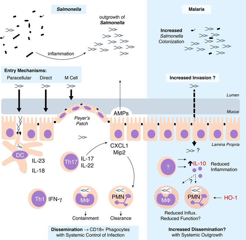
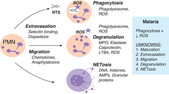
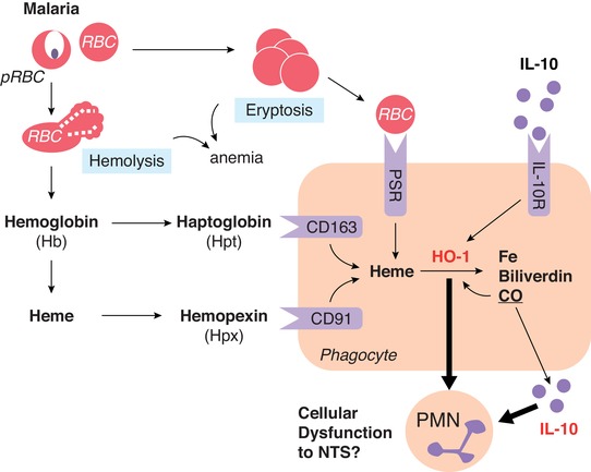
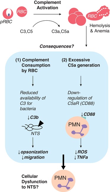
Invasive bacterial disease is well described in immunocompromised hosts, including those with malaria infection. One bacterial infection frequently observed in children with Plasmodium falciparum infection is nontyphoidal salmonella (NTS) infection, in which a typically intestinal infection becomes systemic with serious, often fatal, consequences. In this review, we consider the role of malaria-induced immunoregulatory responses in tipping the balance from tissue homeostasis during malaria infection to risk of invasive NTS. Also, neutrophils are crucial in the clearance of NTS but their ability to mount an oxidative burst and kill intracellular Salmonella is severely compromised during, and for some time after, an acute malaria infection. Here, we summarize the evidence linking malaria and invasive NTS infections; describe the role of neutrophils in clearing NTS infections; review evidence for neutrophil dysfunction in malaria infections; and explore roles of heme oxygenase-1, IL-10, and complement in mediating this dysfunction. Finally, given the epidemiological evidence that low density, subclinical malaria infections pose a risk for invasive NTS infections, we consider whether the high prevalence of such infections might underlie the very high incidence of invasive bacterial disease across much of sub-Saharan Africa.

Keywords: IL-10; anemia; heme oxygenase-1; malaria; neutrophil; salmonella; sepsis.


Figures

Figure 1
Figure 1
NTS intestinal immune response. NTS is a fecal‐oral pathogen, which thrives in the inflamed intestine. Tissue invasion in the distal intestine can be via direct invasion, uptake by M cells, or through paracellular spaces. Uptake by dendritic cells can initiate inflammation through the IL‐23/IL‐17 axis. Th17 cells promote neutrophil influx via the induction of neutrophil chemokines. NTS is able to persist within the Salmonella‐containing vacuole of macrophages, whereas neutrophils are efficient at NTS clearance. Systemic dissemination to draining lymph nodes is through CD18+ phagocytes. During experimental malaria, NTS colonization resistance is lowered, although it is unclear if there is an increase in tissue invasion. Regardless, inflammation (with reduced PMN influx) is reduced due to increased IL‐10 concentrations. However, the role of IL‐10, and potentially HO‐1, on intestinal neutrophil function and role for increased systemic dissemination are unclear
Figure 2
Figure 2
Neutrophil function. PMN extravasation is initiated by inflammatory mediators, which stimulate the upregulation of adhesion molecules, such as selectins, on endothelial cells allowing tethering and rolling of PMN. Chemokine gradients cause PMNs to “crawl” toward the site of infection until an endothelial junction is reached where diapedesis can occur. At the site of infection, neutrophils have numerous functions including phagocytosis, degranulation, and NETosis. Phagocytosis involves the ingestion of a pathogen into a phagolysosome containing granule proteins and ROS, to disable and digest internalized pathogens. Degranulation can also occur extracellularly where granular proteins, such as MPO and elastase, and ROS can be released from PMNs. A novel function of PMNs is NETosis, which is the release of NETs. These decondensed chromatin fibers decorated with histones and granular proteins trap and kill pathogens to prevent further dissemination. It is known that during malaria infection, neutrophils maintain the ability to phagocytose NTS; however, PMNs display impaired ROS production. Mechanisms still to be investigated include: maturation, extravasation, migration, degranulation, and NETosis
Figure 3
Figure 3
Anemia, HO‐1, and IL‐10. During malaria infection, anemia can occur through direct lysis of parasitized RBCs (hemolysis) or through eryptosis of uninfected RBCs—which are taken up by phagocytes through the phosphatidylserine receptor (PSR). Hemolysis results in release of hemoglobin (Hb), which can be further degraded to free heme. Serum scavenger proteins haptoglobin (Hpt) and hemopexin (Hpx) bind Hb and heme before uptake by phagocytes through CD163 and CD91, respectively. Intracellular heme is then degraded to iron, biliverdin, and carbon monoxide via the enzyme HO‐2 or the inducible isoform, HO‐1. HO‐1 can also be induced by IL‐10 receptor signaling. The impact of IL‐10 and HO‐1 during malarial anemia and hemolysis on phagocyte function remains poorly defined
Figure 4
Figure 4
Complement depletion. During malaria infection, systemic complement activation occurs due to deposition on both infected and uninfected RBCs. This results in reduced concentrations of circulating C3 and C5 and generation of anaphylatoxins (C3a and C5a). The consequences that may impact PMN function are 2‐fold; (1) a reduction in C3 available for deposition on extracellular NTS leading to opsonization and migration and/or (2) excessive C5a reducing CD88 (C5aR) on neutrophils leading to reduced ROS and cytokine production. Additional work is needed to clarify the impact of complement activation and subsequent depletion during malaria on risk to NTS bacteremia

References

    1. Shankar‐Hari M, Phillips G, Levy M, et al. Assessment of definition and clinical criteria for septic shock: for the third international consensus definitions for sepsis and septic shock (Sepsis‐3). JAMA. 2016;315:775–787. - PMC - PubMed
    1. Bone RC, Balk RA, Cerra FB, et al. Definitions for sepsis and organ failure and guidelines for the use of innovative therapies in sepsis. American College of Chest Physicians/Society of Critical Care Medicine. Chest. 1992;101:1644–1655. - PubMed
    1. Angus DC, van der Poll T. Severe sepsis and septic shock. N Engl J Med. 2013;369:840–851. - PubMed
    1. Antonelli M. Sepsis and septic shock: pro‐inflammatory or anti‐inflammatory state. J Chemother. 1999;11:536–540. - PubMed
    1. Tanaka H, Sugimoto H, Yoshioka T, Sugimoto T. Role of granulocyte elastase in tissue injury in patients with septic shock complicated by multiple‐organ failure. Ann Surg. 1991;213:81–85. - PMC - PubMed

Publication types