. 2019 Jun;60(6):687-694.
doi: 10.1165/rcmb.2018-0179OC.

Pulmonary Neuroendocrine Cells Secrete γ-Aminobutyric Acid to Induce Goblet Cell Hyperplasia in Primate Models

Affiliations

Pulmonary Neuroendocrine Cells Secrete γ-Aminobutyric Acid to Induce Goblet Cell Hyperplasia in Primate Models

Juliana Barrios et al. Am J Respir Cell Mol Biol. 2019 Jun.

Abstract

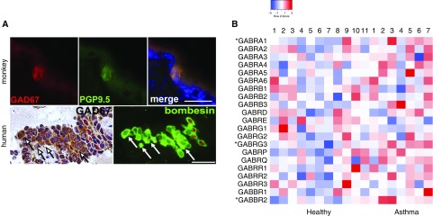
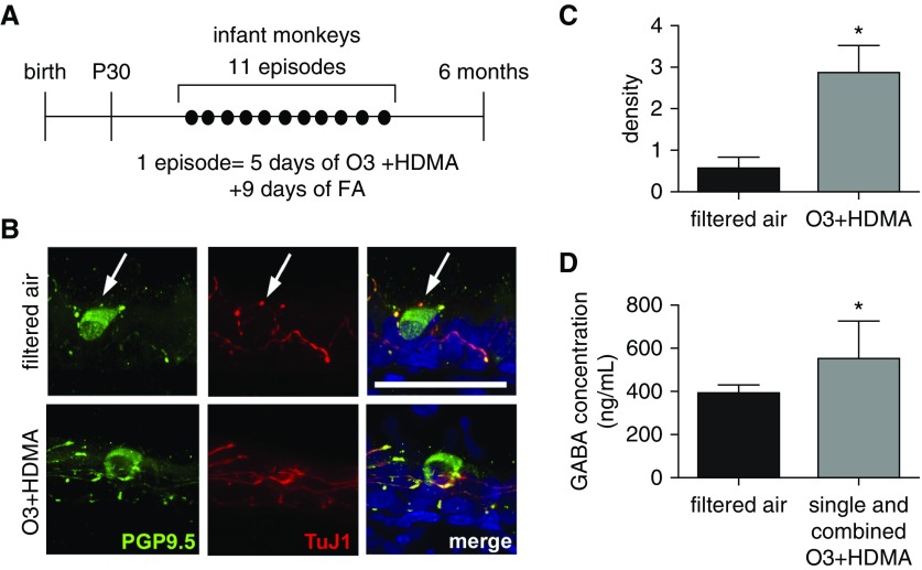
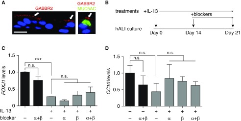
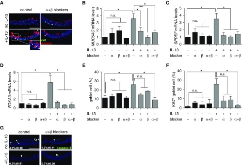
Mucus overproduction is a major contributor to morbidity and mortality in asthma. Mucus overproduction is induced by orchestrated actions of multiple factors that include inflammatory cytokines and γ-aminobutyric acid (GABA). GABA is produced only by pulmonary neuroendocrine cells (PNECs) in the mouse lung. Recent studies in a neonatal mouse model of allergic inflammation have shown that PNECs play an essential role in mucus overproduction by GABA hypersecretion. Whether PNECs mediate dysregulated GABA signaling for mucus overproduction in asthma is unknown. In this study, we characterized the cellular source of GABA in the lungs of nonhuman primates and humans and assessed GABA secretion and signaling in primate disease models. We found that like in mice, PNECs were the major source of GABA in primate lungs. In addition, an infant nonhuman primate model of asthma exhibited an increase in GABA secretion. Furthermore, subjects with asthma had elevated levels of expression of a subset of GABA type α (GABAα) and type β (GABAβ) receptors in airway epithelium compared with those of healthy control subjects. Last, employing a normal human bronchial epithelial cell model of preinduced mucus overproduction, we showed pharmaceutical blockade of GABAα and GABAβ receptor signaling reversed the effect of IL-13 on MUC5AC gene expression and goblet cell proliferation. Together, our data demonstrate an evolutionarily conserved intraepithelial GABA signaling that, in concert with IL-13, plays an essential role in mucus overproduction. Our findings may offer new strategies to ameliorate mucus overproduction in patients with asthma by targeting PNEC secretion and GABA signaling.

Keywords: asthma; goblet cell hyperplasia; mucus overproduction; pulmonary neuroendocrine cell; γ-aminobutyric acid.


Figures

Figure 1.
Figure 1.
Airway epithelial production of γ-aminobutyric acid (GABA) in primate lungs and GABA receptors in healthy humans and humans with asthma. (A) Representative glutamate decarboxylase 67 (GAD67) expression in pulmonary neuroendocrine cells (PNECs). Histological sections of the lung in rhesus monkeys (6 mo old) and infant human donors were double stained for GAD67 and PNEC markers PGP9.5 (protein gene product 9.5) and bombesin, respectively. *Bombesin+ PNECs that are GAD67. Arrows mark nonspecific labeling by the GAD67 antibody in human lung mesenchyme. For quantification, 10 tissue sections from six monkeys were stained, and a total of 129 PNECs were all found to express GAD67. For the human lung, five tissue sections from three donor lungs were stained. Of 74 bombesin+ PNECs, 58 were GAD67+. Scale bars: 25 μm. (B) Relative GABA receptor gene expression in airway epithelium of healthy donors (n = 11) and donors with asthma (n = 7). Data were collected from microarray results of airway epithelial brushing samples deposited in the Gene Expression Omnibus GSE4302 public dataset (contact contributor, P. G. Woodruff). Each column represents one donor. *Genes with statistically significant increases in expression in asthmatic samples.
Figure 2.
Figure 2.
PNEC hyperinnervation and GABA hypersecretion in a nonhuman primate model of early-life exposure. (A) Experimental scheme of rhesus monkey ozone (O3) and house dust mite allergen (HDMA) exposure during the first 6 months after birth. Control animals were exposed to filtered air (FA). Lungs and serum samples were harvested at 6 months of age. (B) Representative Z-stack confocal images of PNEC innervation in 6-month-old rhesus monkeys with and without O3 + HDMA exposure. PNEC innervation was assessed by double staining of proximal lung sections (25 μm in thickness) for PGP9.5 and neuron-specific β-tubulin III using a TuJ1 antibody. Arrows mark PNECs. Scale bar: 50 μm. (C) Quantification of PNEC innervation in infant rhesus monkeys with and without O3 and HDMA exposure. The density of nerves surrounding PNECs was calculated by normalizing TuJ1-immunoreactive area to the number of PNECs. At least five single PNECs and PNEC clusters from three lungs in each group were measured. (D) Serum concentrations of GABA in control animals (n = 7) and rhesus monkeys (n = 10) that were exposed to O3, HDMA, and combined O3 + HDMA. Data presented in C and D represent mean ± SEM. *P < 0.05.
Figure 3.
Figure 3.
GABAα and GABAβ signaling has no effect on baseline and IL-13–induced differentiation of air–liquid interface (hALI) cultures of normal human bronchial epithelial (NHBE) cells. (A) GABAβ receptor (GABBR2) expression in ALI cultures at Day 14 of differentiation by double staining for GABBR2 and MUC5AC. Nuclei were stained by DAPI. Arrows mark GABBR2 expression in MUC5AC+ goblet cells and other epithelial cell types. Scale bar: 50 μm. (B) Scheme of the treatment with GABA receptor blockers in control and IL-13–stimulated ALI cultures. IL-13 (10 ng/ml) stimulation started at Day 0. Treatment with single and combined GABAα receptor blocker (50 μM picrotoxin) and GABAβ receptor blocker (1 μM CGP55845) started at Day 14. (C and D) Cultures were analyzed at Day 21 for the gene expression of a ciliated cell marker (FOXJ1) (C) and a club cell marker (CC10) (D). Data represent mean ± SEM of three independent experiments of NHBE cells from each donor and a total of three donors for each condition. ***P < 0.001. n.s. = not significant.
Figure 4.
Figure 4.
GABA is required for IL-13–induced goblet cell hyperplasia in ALI cultures of NHBE cells. (A) Representative images of double staining for Ki-67 and MUC5AC in Day 21 ALI cultures of NHBE cells with and without the treatment of IL-13 and combined GABAα and GABAβ receptor blockers. Arrowheads mark MUC5AC+Ki-67+ cells. The inset shows an enlarged image of double-positive cells in IL-13–treated cultures. (BD) MUC5AC, SPDEF (SAM pointed domain-containing Ets transcription factor), and FOXA3 gene expression in Day 21 ALI cultures of NHBE cells under different culture conditions. (E) Quantification of the percentages of MUC5AC+ goblet cells in Day 21 ALI cultures under different culture conditions. (F) Goblet cell proliferation in Day 21 ALI cultures under different culture conditions. Proliferation was quantified by normalizing the number of MUC5AC+Ki-67+ cells to the total MUC5AC+ goblet cells. More than 500 cells from each donor were counted for each condition. (G) Representative images of staining for cleaved caspase 3 in Day 21 ALI cultures with and without the treatment of IL-13 and combined GABAα and GABAβ receptor blockers. Data in BD represent mean ± SEM of three independent experiments in triplicates for each donor, three donors in total. *P < 0.05 and **P < 0.01. Arrowheads mark caspase 3+ cells. Scale bars: 50 μm.

References

    1. Fahy JV, Dickey BF. Airway mucus function and dysfunction. N Engl J Med. 2010;363:2233–2247. - PMC - PubMed
    1. Maddox L, Schwartz DA. The pathophysiology of asthma. Annu Rev Med. 2002;53:477–498. - PubMed
    1. Stern DA, Morgan WJ, Halonen M, Wright AL, Martinez FD. Wheezing and bronchial hyper-responsiveness in early childhood as predictors of newly diagnosed asthma in early adulthood: a longitudinal birth-cohort study. Lancet. 2008;372:1058–1064. - PMC - PubMed
    1. Aven L, Paez-Cortez J, Achey R, Krishnan R, Ram-Mohan S, Cruikshank WW, et al. An NT4/TrkB-dependent increase in innervation links early-life allergen exposure to persistent airway hyperreactivity. FASEB J. 2014;28:897–907. - PMC - PubMed
    1. Herring MJ, Putney LF, St George JA, Avdalovic MV, Schelegle ES, Miller LA, et al. Early life exposure to allergen and ozone results in altered development in adolescent rhesus macaque lungs. Toxicol Appl Pharmacol. 2015;283:35–41. - PMC - PubMed

Publication types