. 2018 Nov 6;7(21):e009098.
doi: 10.1161/JAHA.118.009098.

Elevated Inflammatory Plasma Biomarkers in Patients With Fabry Disease: A Critical Link to Heart Failure With Preserved Ejection Fraction

Affiliations

Elevated Inflammatory Plasma Biomarkers in Patients With Fabry Disease: A Critical Link to Heart Failure With Preserved Ejection Fraction

Haran Yogasundaram et al. J Am Heart Assoc. .

Abstract

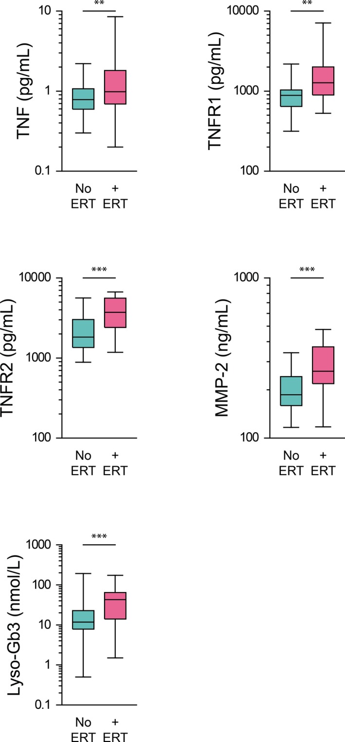
Background Because systemic inflammation and endothelial dysfunction lead to heart failure with preserved ejection fraction, we characterized plasma levels of inflammatory and cardiac remodeling biomarkers in patients with Fabry disease ( FD ). Methods and Results Plasma biomarkers were studied in multicenter cohorts of patients with FD (n=68) and healthy controls (n=40). Plasma levels of the following markers of inflammation and cardiac remodeling were determined: tumor necrosis factor ( TNF ), TNF receptor 1 ( TNFR 1) and 2 ( TNFR 2), interleukin-6, matrix metalloprotease-2 ( MMP -2), MMP -8, MMP -9, galectin-1, galectin-3, B-type natriuretic peptide ( BNP ), midregional pro-atrial natriuretic peptide ( MR -pro ANP ), and globotriaosylsphingosine. Clinical profile, cardiac magnetic resonance imaging, and echocardiogram were reviewed and correlated with biomarkers. Patients with FD had elevated plasma levels of BNP , MR -pro ANP , MMP -2, MMP -9, TNF , TNFR 1, TNFR 2, interleukin-6, galectin-1, globotriaosylsphingosine, and analogues. Plasma TNFR 2, TNF , interleukin-6, MMP -2, and globotriaosylsphingosine were elevated in FD patients with left ventricular hypertrophy, whereas diastolic dysfunction correlated with higher BNP , MR -pro ANP , and MMP -2 levels. Patients with late gadolinium enhancement on cardiac magnetic resonance imaging had greater levels of BNP , MR -pro ANP , TNFR 1, TNFR 2, and MMP -2. Plasma BNP , MR -pro ANP , MMP -2, MMP -8, TNF , TNFR 1, TNFR 2, galectin-1, and galectin-3 were elevated in patients with renal dysfunction. Patients undergoing enzyme replacement therapy who have more severe disease had higher MMP -2, TNF , TNFR 1, TNFR 2, and globotriaosylsphingosine analogue levels. Conclusions Inflammatory and cardiac remodeling biomarkers are elevated in FD patients and correlate with disease progression. These features are consistent with a phenotype dominated by heart failure with preserved ejection fraction and suggest a key pathogenic role of systemic inflammation in FD .

Keywords: Fabry disease; biomarkers; heart failure with preserved ejection fraction; hypertrophy; inflammation.


Figures

Figure 1
Figure 1
Examples of ion chromatograms for lysoglobotriaosylceramide (Lyso‐Gb3), its 6 analogues, and Lyso‐Gb3‐Gly (used as the internal standard) detected in plasma from a Fabry patient. The (+H2O2) analogue has 2 structural isomers with retention times of 3.29 and 4.57 minutes. The areas of these peaks were added together for computation results. cps indicates counts per second.
Figure 2
Figure 2
Plasma levels of cardiac remodeling biomarkers and Lyso‐Gb3 in cohorts of patients with FD (n=68) and healthy controls (n=40). Biomarkers BNP, MR‐proANP, galectin‐1, galectin‐3, and Lyso‐Gb3 are significantly elevated in the FD cohort relative to healthy controls. BNP indicates B‐type natriuretic peptide; FD, Fabry disease; HC, healthy controls; Lyso‐Gb3, lysoglobotriaosylceramide; MR‐proANP, midregional pro–atrial natriuretic peptide. *P<0.05; **P<0.01; ***P<0.001.
Figure 3
Figure 3
Plasma levels of inflammatory biomarkers and selected matrix metalloproteases in cohorts of FD (n=68) and healthy controls (n=40). TNF, IL‐6, TNFR1, and TNFR2 are significantly elevated in the FD cohort relative to healthy controls, without significant elevation in CRP. In addition, MMP‐2 and MMP‐9 are also significantly elevated in the FD cohort relative to healthy controls, although no difference was observed for MMP‐8. CRP, C‐reactive peptide; FD, Fabry disease; HC, healthy controls; IL, interleukin; MMP, matrix metalloprotease; TNF, tumor necrosis factor; TNFR, TNF receptor. *P<0.05; **P<0.01; ***P<0.001.
Figure 4
Figure 4
Receiver operating characteristic (ROC) curve demonstrating the performance of Lyso‐Gb3 and Lyso‐Gb3 with analogues for the prediction of FD (n=40 healthy controls; n=68 FD patients). Lyso‐Gb3, the gold standard, had excellent performance (AUC=0.998), and Lyso‐Gb3 with analogues had an AUC=0.994. AUC indicates area under the curve; FD, Fabry disease; Lyso‐Gb3, lysoglobotriaosylceramide.
Figure 5
Figure 5
Plasma levels of biomarkers in FD patients (n=68) with (n=41) and without (n=27) LVH via imaging criteria. TNFR2, TNF, IL‐6, MMP‐2, and Lyso‐Gb3 are significantly higher in FD patients with LVH than in those without LVH. FD indicates Fabry disease; IL, interleukin; LVH, left ventricular hypertrophy (left ventricular mass index ≥85 g/m2 in male and ≥81 g/m2 in female patients); Lyso‐Gb3, lysoglobotriaosylceramide; MMP, matrix metalloprotease; TNF, tumor necrosis factor; TNFR, TNF receptor. *P<0.05; **P<0.01.
Figure 6
Figure 6
Plasma levels of biomarkers in FD patients (n=65) with (n=30) and without (n=35) late gadolinium enhancement (LGE) on cardiac MRI. BNP, MR‐proANP, TNFR1, TNFR2, and MMP‐2 are elevated in FD patients with LGE. BNP indicates B‐type natriuretic peptide; FD, Fabry disease; LGE, late gadolinium enhancement; MMP, matrix metalloprotease; MRI, magnetic resonance imaging; MR‐proANP, midregional pro–atrial natriuretic peptide; TNFR, tumor necrosis factor receptor. **P<0.01; ***P<0.001.
Figure 7
Figure 7
A, Plasma levels of biomarkers in FD patients (n=43) with (n=17) and without diastolic dysfunction (n=26) per echocardiography. BNP, MR‐proANP, and MMP‐2 are significantly higher in FD patients with diastolic dysfunction than in those without diastolic dysfunction. Diastolic dysfunction could not be assessed in some patients due to arrhythmia. B, Correlation plot of MR‐proANP vs maximum LA size index (R 2=0.44, P<0.001). MR‐proANP and maximum LA size index are positively correlated, as increasing LA size is known to cause atrial cardiomyocytes to release ANP. BNP indicates B‐type natriuretic peptide; DD, diastolic dysfunction; FD, Fabry disease; LA, left atrial; MMP, matrix metalloprotease; MR‐proANP, midregional pro–atrial natriuretic peptide. *P<0.05; **P<0.01.
Figure 8
Figure 8
Plasma levels of biomarkers in FD patients (n=68) with (n=18) and without significant chronic kidney disease (n=50). BNP, MR‐proANP, TNF, TNFR1, TNFR2, MMP‐2, MMP‐8, galectin‐1, and galectin‐3 are significantly elevated in FD patients with CKD. BNP indicates B‐type natriuretic peptide; CKD, chronic kidney disease (estimated glomerular filtration rate <60 mL/[min·1.73 m2]); FD, Fabry disease; MMP, matrix metalloprotease; MR‐proANP, midregional pro–atrial natriuretic peptide; TNF, tumor necrosis factor; TNFR, TNF receptor. *P<0.05; **P<0.01; ***P<0.001.
Figure 9
Figure 9
Plasma levels of biomarkers in FD patients (n=68), in cohorts of those not receiving ERT (n=31) and those who qualify for and are receiving ERT (n=37). TNF, TNFR1, TNFR2, MMP‐2, and Lyso‐Gb3 are elevated in FD patients undergoing ERT relative to those not receiving ERT. ERT indicates enzyme replacement therapy; FD, Fabry disease; Lyso‐Gb3, lysoglobotriaosylceramide; MMP, matrix metalloprotease; TNF, tumor necrosis factor; TNFR, TNF receptor. **P<0.01; ***P<0.001.
Figure 10
Figure 10
Plasma levels of biomarkers in FD patients (n=68) of male (n=34) and female (n=34) sex. TNF, TNFR1, TNFR2, MMP‐2, and Lyso‐Gb3 are elevated in male FD patients relative to female patients. F indicates female; FD, Fabry disease; Lyso‐Gb3, lysoglobotriaosylceramide; M, male; MMP, matrix metalloprotease; TNF, tumor necrosis factor; TNFR, TNF receptor. *P<0.05; **P<0.01; ***P<0.001.

References

    1. Eng CM, Resnick‐Silverman LA, Niehaus DJ, Astrin KH, Desnick RJ. Nature and frequency of mutations in the alpha‐galactosidase A gene that cause Fabry disease. Am J Hum Genet. 1993;53:1186–1197. - PMC - PubMed
    1. Niemann M, Rolfs A, Stork S, Bijnens B, Breunig F, Beer M, Ertl G, Wanner C, Weidemann F. Gene mutations versus clinically relevant phenotypes: lyso‐Gb3 defines Fabry disease. Circ Cardiovasc Genet. 2014;7:8–16. - PubMed
    1. Lin HY, Chong KW, Hsu JH, Yu HC, Shih CC, Huang CH, Lin SJ, Chen CH, Chiang CC, Ho HJ, Lee PC, Kao CH, Cheng KH, Hsueh C, Niu DM. High incidence of the cardiac variant of Fabry disease revealed by newborn screening in the Taiwan Chinese population. Circ Cardiovasc Genet. 2009;2:450–456. - PubMed
    1. Inoue T, Hattori K, Ihara K, Ishii A, Nakamura K, Hirose S. Newborn screening for Fabry disease in Japan: prevalence and genotypes of Fabry disease in a pilot study. J Hum Genet. 2013;58:548–552. - PubMed
    1. Gambarin FI, Disabella E, Narula J, Diegoli M, Grasso M, Serio A, Favalli V, Agozzino M, Tavazzi L, Fraser AG, Arbustini E. When should cardiologists suspect Anderson‐Fabry disease? Am J Cardiol. 2010;106:1492–1499. - PubMed

Publication types