. 2018 Nov 9;123(11):e35-e47.
doi: 10.1161/CIRCRESAHA.118.313283.

Macrophage Inflammation, Erythrophagocytosis, and Accelerated Atherosclerosis in Jak2 V617F Mice

Affiliations

Macrophage Inflammation, Erythrophagocytosis, and Accelerated Atherosclerosis in Jak2 V617F Mice

Wei Wang et al. Circ Res. .

Abstract

Rationale: The mechanisms driving atherothrombotic risk in individuals with JAK2 V617F ( Jak2 VF) positive clonal hematopoiesis or myeloproliferative neoplasms are poorly understood.

Objective: The goal of this study was to assess atherosclerosis and underlying mechanisms in hypercholesterolemic mice with hematopoietic Jak2 VF expression.

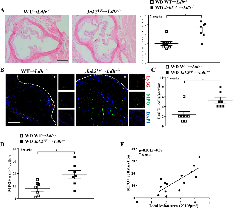
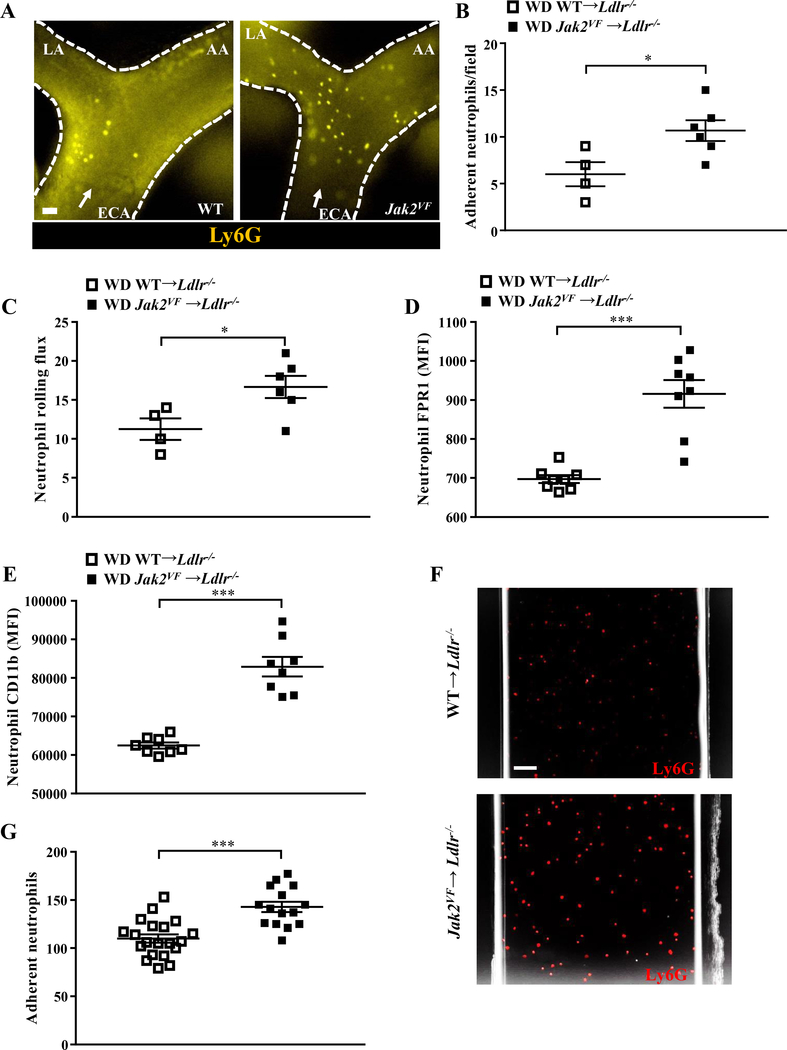
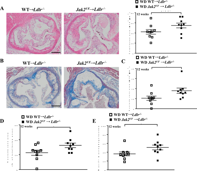
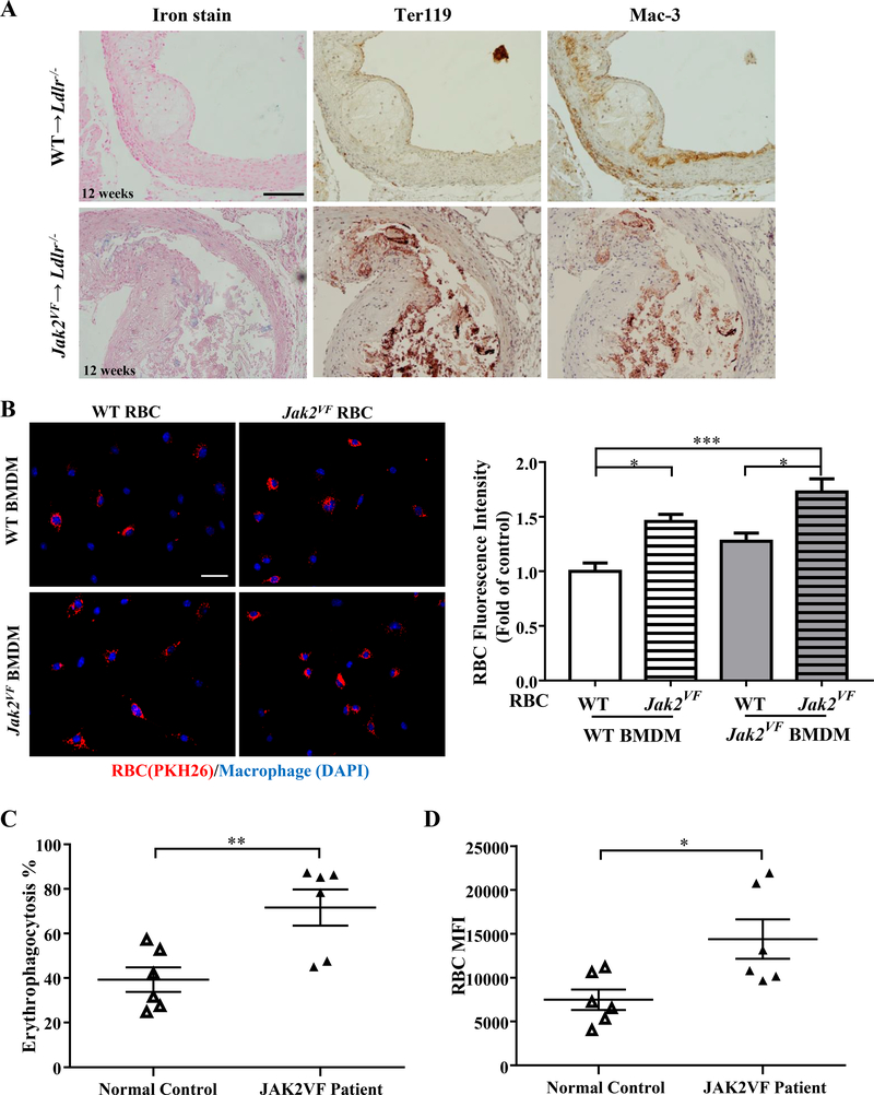
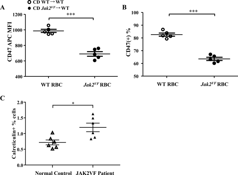
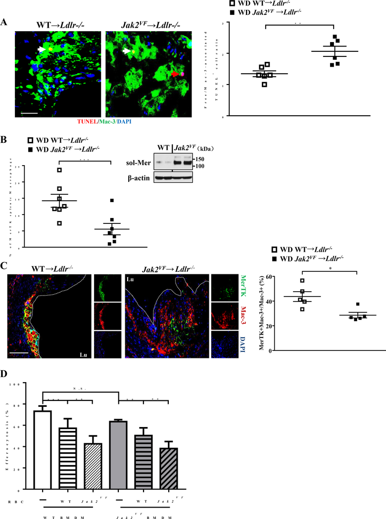
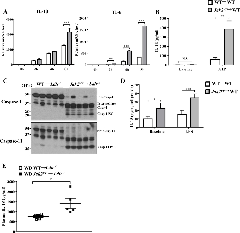
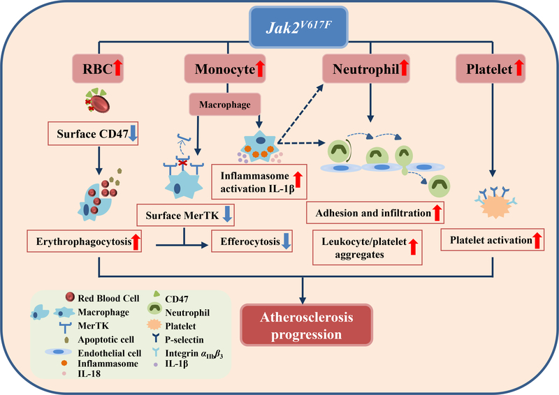
Methods and results: Irradiated low-density lipoprotein receptor knockout ( Ldlr-/-) mice were transplanted with bone marrow from wild-type or Jak2 VF mice and fed a high-fat high-cholesterol Western diet. Hematopoietic functions and atherosclerosis were characterized. After 7 weeks of Western diet, Jak2 VF mice showed increased atherosclerosis. Early atherosclerotic lesions showed increased neutrophil adhesion and content, correlating with lesion size. After 12 weeks of Western diet, Jak2 VF lesions showed increased complexity, with larger necrotic cores, defective efferocytosis, prominent iron deposition, and costaining of erythrocytes and macrophages, suggesting erythrophagocytosis. Jak2 VF erythrocytes were more susceptible to phagocytosis by wild-type macrophages and showed decreased surface expression of CD47, a "don't-eat-me" signal. Human JAK2VF erythrocytes were also more susceptible to erythrophagocytosis. Jak2 VF macrophages displayed increased expression and production of proinflammatory cytokines and chemokines, prominent inflammasome activation, increased p38 MAPK (mitogen-activated protein kinase) signaling, and reduced levels of MerTK (c-Mer tyrosine kinase), a key molecule mediating efferocytosis. Increased erythrophagocytosis also suppressed efferocytosis.

Conclusions: Hematopoietic Jak2 VF expression promotes early lesion formation and increased complexity in advanced atherosclerosis. In addition to increasing hematopoiesis and neutrophil infiltration in early lesions, Jak2 VF caused cellular defects in erythrocytes and macrophages, leading to increased erythrophagocytosis but defective efferocytosis. These changes promote accumulation of iron in plaques and increased necrotic core formation which, together with exacerbated proinflammatory responses, likely contribute to plaque instability.

Keywords: atherosclerosis; erythrocytes; inflammasomes; inflammation; macrophages.


Conflict of interest statement

DISCLOSURE

The authors disclose no conflict of interest.

Figures

Figure 1:
Figure 1:. Increased early atherosclerotic lesions and neutrophil infiltration in Jak2VF mice.
(A) Representative H&E-stained aortic root lesions and quantification of total lesion area of female Ldlr−/− recipients after 7 weeks of WD. Mann-Whitney U test. Scale bar, 500μm. (B) Representative immunofluorescence images of MPO (Green) and Ly6G (Red) with DAPI (Blue) of aortic root lesions from female mice fed WD for 7 weeks. Mann-Whitney U test. Scale bar,100μm. (C) Quantification of Ly6G positive cells (Mann-Whitney U test) and (D) MPO positive cells in the lesions. Unpaired t test. (E) Correlation between MPO+ neutrophils and total lesion size after 7 weeks of WD. *p<0.05, **p<0.01. Spearman correlation test.
Figure 2:
Figure 2:. Increased rolling and adhesion of neutrophils in Jak2VF mice.
Female Ldlr−/− recipients were fed WD for 5 weeks. (A) Representative image of epifluorescence intravital microscopy of the carotid artery showing interaction of Ly6G-stained neutrophils with the arterial vessel wall. ECA, external carotid artery; LA, lingual artery; AA, auricular artery. Arrow indicates flow direction. Scale bar, 50μm. (B) Quantification of Ly6G-stained neutrophil adhesion in the carotid artery by intravital microscopy. Mann-Whitney U test. (C) Neutrophil rolling flux was assessed by intravital microscopy. Expression of (D) FPR1 and (E) CD11b MFI on neutrophils were measured by flow cytometry. Mann-Whitney U test. (F,G) Flow chamber assays for neutrophil adhesion using equal number of neutrophils. (F) Representative images and (G) quantification of adhesion. Mann-Whitney U test. Scale bar, 50μm. *p<0.05, ***p<0.001.
Figure 3:
Figure 3:. Increased necrotic core in advanced lesions of Jak2VF mice.
(A) Representative H&E-stained aortic root lesions and quantification of total lesion area of female Ldlr−/− recipients after 12 weeks of WD. Unpaired t test. Scale bar, 500μm. (B) Representative massion trichrome stain images of lesions. Scale bar, 500μm. (C) Quantification of necrotic core area and (D) as a percentage of total lesion area of female Ldlr−/− recipients after 12 weeks WD. Mann-Whitney U test. (E) Quantification of Mac-3+ macrophage in the lesions of female mice WD-fed for 12 weeks. Mann-Whitney U test. *p<0.05.
Figure 4:
Figure 4:. Jak2VF mice displayed marked increase in erythrophagocytosis.
(A) Representative images of iron staining, immunohistochemistry staining of red blood cell marker Ter119 and macrophage marker Mac-3 in lesions of female Ldlr−/− recipients after 12 weeks WD. Scale bar, 100μm. (B) Bone marrow derived macrophages (DAPI, Blue) were incubated with 2 million PKH26-labeled erythrocytes (Red) overnight and quantification of relative red blood cells fluorescence intensity. Cells from both male and female mice were used for the assays. Pooled data from 5 independent experiments were used for analysis. Scale bar, 50μm. 2-way ANOVA. (C) Erythrophagocytosis rate (Mann-Whitney U test) and (D) red blood cells MFI of human normal control and JAK2VF patients were measured by flow cytometry. Unpaired t test. *p<0.05, **p<0.01,***p<0.001.
Figure 5:
Figure 5:. Jak2VF mice showed reduced surface CD47 expression in erythrocytes.
Erythrocytes were from chow-fed female mice. (A) Mean fluorescence intensity of anti-CD47 antibody bound to erythrocytes (Unpaired t test) and (B) percentage of CD47hi erythrocytes by flow cytometry. Unpaired t test. (C) Percentage of calreticulin positive erythrocytes in human normal controls and JAK2VF patients. Unpaired t test.*p<0.05, ***p<0.001.
Figure 6:
Figure 6:. Defective efferocytosis was associated with decreased macrophage surface MerTK in Jak2VF mice.
(A) Representative images of advanced lesions (12 weeks WD-fed) in which apoptotic cells were stained by TUNEL (Red), macrophages by Mac-3 (Green) and nuclei by DAPI (Blue). Efferocytosis was assessed as the ratio of free to macrophage-associated TUNEL positive cells. The red arrow depicts free apoptotic cells and the white arrow depicts macrophage associated apoptotic cells. Scale bar, 20μm. Unpaired t test. (B) Percentage of MerTK postive macrophages in total spleen cells as determined by flow cytometry and western blot of soluble MerTK levels in cultured media of splenic macrophages. Unpaired t test. (C) Representative single (small panel) or merged fluorescence images (large panel) of Mac-3 (Red), MerTK (Green) or DAPI (Blue) and quantification of the ratio of MerTK/Mac-3 co-positive to Mac-3 positive macrophages. Scale bar, 100μm. Mann-Whitney U test. (D) Bone marrow derived WT or Jak2VF macrophages were treated with or without 5 million WT or Jak2VF erythrocytes in the presence of apoptotic Jurkat cells for 20 hours to assess efferocytosis by fluorescence microscope. Data were the representative of 5 independent experiments. 2-way ANOVA. *p<0.05, **p<0.01, ***p<0.001.
Figure 7:
Figure 7:. Jak2VF myeloid cells displayed enhance inflammasome activation.
(A) Con-A induced peritoneal macrophages were challenged with or without 10ng/ml LPS for the indicated time and qPCR analysis of mRNA level of IL-1β and IL-6. Data were from 5 independent experiments. 1-way ANOVA. (B) ELISA of IL-1β in cultured medium of bone marrow derived macrophage challenged with 10ng/ml LPS for 1 hour followed by 1mM ATP for 3 hours. Baseline was LPS (10ng/ml) only for 4 hours. 1-way ANOVA. (C) Western blot of caspase-1 and caspase-11 cleavage in splenic CD11b+ cells from female recipients WD-fed for 7 weeks. (D) ELISA of IL-1β in cultured medium of CD11b+ cells from female recipients WD-fed for 7 weeks. Cells were treated with or without 1μg/ml LPS for 8h. Data were the representative from 4 independent experiments. 2-way ANOVA. (E) ELISA of plasma IL-18 in female recipients WD-fed for 8 weeks. Unpaired t test. *p<0.05, **p<0.01, ***p<0.001.
Figure 8:
Figure 8:. Schematic model.
Mechanisms underlying increased atherosclerosis in Jak2VF mice.

Comment in

References

    1. Campbell PJ and Green AR. The myeloproliferative disorders. The New England journal of medicine. 2006;355:2452–66. - PubMed
    1. James C, Ugo V, Le Couedic JP, Staerk J, Delhommeau F, Lacout C, Garcon L, Raslova H, Berger R, Bennaceur-Griscelli A, Villeval JL, Constantinescu SN, Casadevall N and Vainchenker W. A unique clonal JAK2 mutation leading to constitutive signalling causes polycythaemia vera. Nature. 2005;434:1144–8. - PubMed
    1. Baxter EJ, Scott LM, Campbell PJ, East C, Fourouclas N, Swanton S, Vassiliou GS, Bench AJ, Boyd EM, Curtin N, Scott MA, Erber WN, Green AR and Cancer Genome P. Acquired mutation of the tyrosine kinase JAK2 in human myeloproliferative disorders. Lancet. 2005;365:1054–61. - PubMed
    1. Kralovics R, Passamonti F, Buser AS, Teo SS, Tiedt R, Passweg JR, Tichelli A, Cazzola M and Skoda RC. A gain-of-function mutation of JAK2 in myeloproliferative disorders. The New England journal of medicine. 2005;352:1779–90. - PubMed
    1. Zhao R, Xing S, Li Z, Fu X, Li Q, Krantz SB and Zhao ZJ. Identification of an acquired JAK2 mutation in polycythemia vera. The Journal of biological chemistry. 2005;280:22788–92. - PMC - PubMed

Publication types

MeSH terms

LinkOut - more resources