

Regulation of cardiomyocyte maturation during critical perinatal window
- PMID: 30571853
- PMCID: PMC7682257
- DOI: 10.1113/JP276754
Regulation of cardiomyocyte maturation during critical perinatal window
Abstract
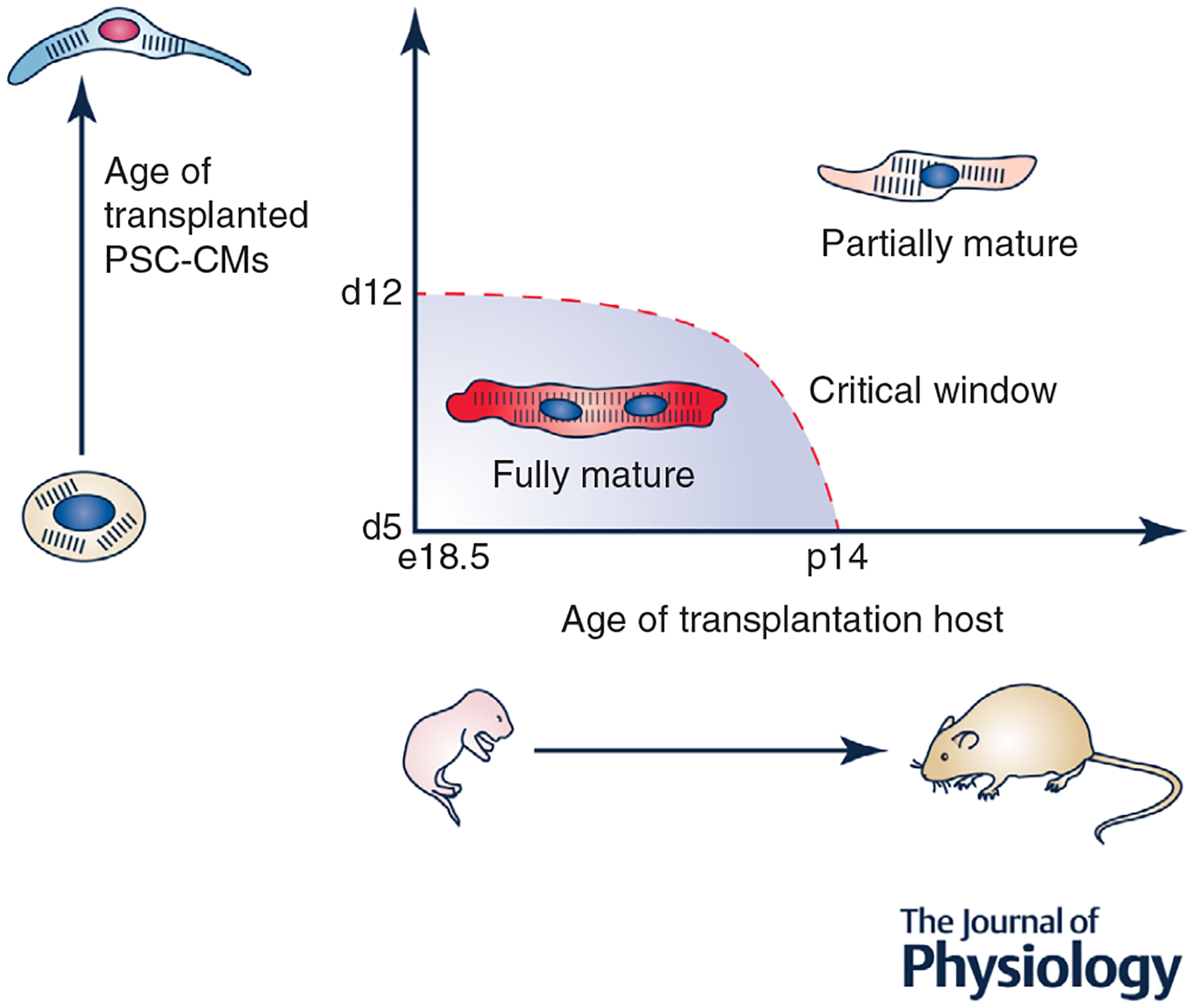
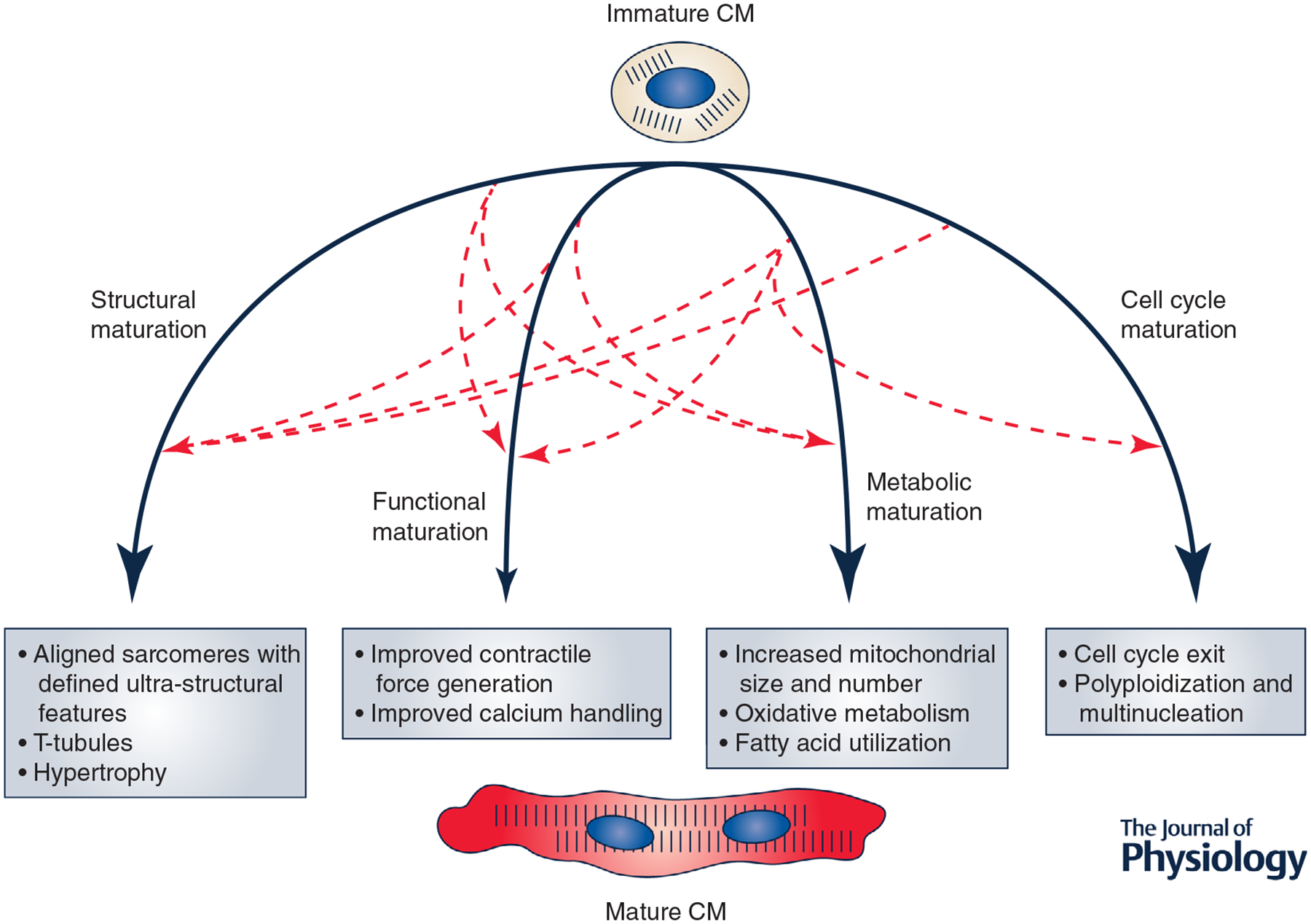
A primary limitation in the use of pluripotent stem cell-derived cardiomyocytes (PSC-CMs) for both patient health and scientific investigation is the failure of these cells to achieve full functional maturity. In vivo, cardiomyocytes undergo numerous adaptive structural, functional and metabolic changes during maturation. By contrast, PSC-CMs fail to fully undergo these developmental processes, instead remaining arrested at an embryonic stage of maturation. There is thus a significant need to understand the biological processes underlying proper CM maturation in vivo. Here, we discuss what is known regarding the initiation and coordination of CM maturation. We postulate that there is a critical perinatal window, ranging from embryonic day 18.5 to postnatal day 14 in mice, in which the maturation process is exquisitely sensitive to perturbation. While the initiation mechanisms of this process are unknown, it is increasingly clear that maturation proceeds through interconnected regulatory circuits that feed into one another to coordinate concomitant structural, functional and metabolic CM maturation. We highlight PGC1α, SRF and the MEF2 family as transcription factors that may potentially mediate this cross-talk. We lastly discuss several emerging technologies that will facilitate future studies into the mechanisms of CM maturation. Further study will not only produce a better understanding of its key processes, but provide practical insights into developing a robust strategy to produce mature PSC-CMs.
Keywords: cardiac; cardiomyocyte; disease modeling; maturation; pluripotent stem cells; regenerative medicine.
© 2018 The Authors. The Journal of Physiology © 2018 The Physiological Society.
Conflict of interest statement
Competing interests
The authors declare no competing interests with regard to this manuscript.
Figures

Similar articles
-
Trajectory reconstruction identifies dysregulation of perinatal maturation programs in pluripotent stem cell-derived cardiomyocytes.Cell Rep. 2023 Apr 25;42(4):112330. doi: 10.1016/j.celrep.2023.112330. Epub 2023 Apr 3. Cell Rep. 2023. PMID: 37014753 Free PMC article.
-
Qualitative transcriptional signatures for evaluating the maturity degree of pluripotent stem cell-derived cardiomyocytes.Stem Cell Res Ther. 2019 Mar 29;10(1):113. doi: 10.1186/s13287-019-1205-1. Stem Cell Res Ther. 2019. PMID: 30925936 Free PMC article.
-
MicroRNA-mediated maturation of human pluripotent stem cell-derived cardiomyocytes: Towards a better model for cardiotoxicity?Food Chem Toxicol. 2016 Dec;98(Pt A):17-24. doi: 10.1016/j.fct.2016.05.025. Epub 2016 Jun 3. Food Chem Toxicol. 2016. PMID: 27265266 Review.
-
Neonatal Transplantation Confers Maturation of PSC-Derived Cardiomyocytes Conducive to Modeling Cardiomyopathy.Cell Rep. 2017 Jan 10;18(2):571-582. doi: 10.1016/j.celrep.2016.12.040. Cell Rep. 2017. PMID: 28076798 Free PMC article.
-
Mitochondria and metabolic transitions in cardiomyocytes: lessons from development for stem cell-derived cardiomyocytes.Stem Cell Res Ther. 2021 Mar 12;12(1):177. doi: 10.1186/s13287-021-02252-6. Stem Cell Res Ther. 2021. PMID: 33712058 Free PMC article. Review.
Cited by
-
PGC1/PPAR drive cardiomyocyte maturation at single cell level via YAP1 and SF3B2.Nat Commun. 2021 Mar 12;12(1):1648. doi: 10.1038/s41467-021-21957-z. Nat Commun. 2021. PMID: 33712605 Free PMC article.
-
Metabolic Remodeling during Early Cardiac Lineage Specification of Pluripotent Stem Cells.Metabolites. 2023 Oct 17;13(10):1086. doi: 10.3390/metabo13101086. Metabolites. 2023. PMID: 37887411 Free PMC article.
-
Pharmacologic therapy for engraftment arrhythmia induced by transplantation of human cardiomyocytes.Stem Cell Reports. 2021 Oct 12;16(10):2473-2487. doi: 10.1016/j.stemcr.2021.08.005. Epub 2021 Sep 9. Stem Cell Reports. 2021. PMID: 34506727 Free PMC article.
-
Advancing Cardiovascular Drug Screening Using Human Pluripotent Stem Cell-Derived Cardiomyocytes.Int J Mol Sci. 2024 Jul 21;25(14):7971. doi: 10.3390/ijms25147971. Int J Mol Sci. 2024. PMID: 39063213 Free PMC article.
-
Dynamic chromatin landscape encodes programs for perinatal transition of cardiomyocytes.Cell Death Discov. 2023 Jan 18;9(1):11. doi: 10.1038/s41420-023-01322-3. Cell Death Discov. 2023. PMID: 36653336 Free PMC article.
References
-
- Agata Y, Hiraishi S, Oguchi K, Misawa H, Horiguchi Y, Fujino N, Yashiro K & Shimada N (1991). Changes in left ventricular output from fetal to early neonatal life. J Pediatr 119, 441–445. - PubMed
-
- Alfar EA, El-Armouche A & Guan K (2018). MicroRNAs in cardiomyocyte differentiation and maturation. Cardiovasc Res 114, 779–781. - PubMed
-
- Anatskaya OV, Sidorenko NV, Beyer TV & Vinogradov AE (2010). Neonatal cardiomyocyte ploidy reveals critical windows of heart development. Int J Cardiol 141, 81–91. - PubMed
Publication types
MeSH terms
Grants and funding
- R01HD086026/HHS | NIH | National Institute of Child Health and Human Development (NICHD)/International
- R01HD086026/National Institute of Child Health and Human Development/International
- R01 HD086026/HD/NICHD NIH HHS/United States
- 18EIA33890038/AHA/American Heart Association-American Stroke Association/United States
- 18EIA33890038/American Heart Association (AHA)/International
LinkOut - more resources
Full Text Sources
Miscellaneous