Review
. 2018 Dec 19;100(6):1292-1311.
doi: 10.1016/j.neuron.2018.11.009.

Microglia in Pain: Detrimental and Protective Roles in Pathogenesis and Resolution of Pain

Affiliations
Review

Microglia in Pain: Detrimental and Protective Roles in Pathogenesis and Resolution of Pain

Gang Chen et al. Neuron. .

Abstract

The previous decade has seen a rapid increase in microglial studies on pain, with a unique focus on microgliosis in the spinal cord after nerve injury and neuropathic pain. Numerous signaling molecules are altered in microglia and contribute to the pathogenesis of pain. Here, we discuss how microglial signaling regulates spinal cord synaptic plasticity in acute and chronic pain conditions with different degrees and variations of microgliosis. We highlight that microglial mediators such as pro- and anti-inflammatory cytokines are powerful neuromodulators that regulate synaptic transmission and pain via neuron-glial interactions. We also reveal an emerging role of microglia in the resolution of pain, in part via specialized pro-resolving mediators including resolvins, protectins, and maresins. We also discuss a possible role of microglia in chronic itch.

Keywords: central sensitization; inflammation; itch; microglia; nerve injury; neuroinflammation; neuropathic pain; specialized pro-resolving mediators; spinal cord; synaptic plasticity.


Figures

Figure-1:
Figure-1:. PubMed search for microglia and neurological diseases.
(A) Number of publications involving microglia in pain, Alzheimer’s disease (AD), Parkinson disease (PD), Huntington disease (HD), multiple sclerosis (MS), stroke, depression, autism, schizophrenia, and anxiety. (B) Number of publications for microglia and pain and microglia and neuropathic pain in last 10 years.
Figure-2:
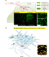
Figure-2:. Nerve injury-induced microgliosis and microglial activation in the spinal cord.
(A) Microgliosis in SDH after nerve injury. (1) Nerve injury (CCI) induces microglial proliferation on the ipsilateral side as revealed in CX3CR1-GFP mice. (2) Nerve injury also induces profound morphological changes of microglia as revealed by CD11b staining with OX-42 antibody. (B) Microglia activation after nerve injury results in the production of microglial mediators, such as TNF, BDNF, IL-1β, and IL-18, which can promote neuropathic pain.
Figure-3:
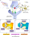
Figure-3:
Schematic illustration of microglial regulations of acute and persistent pain in spinal cord pain circuit via multiple mechanisms, including presynaptic mechanisms and post-synaptic mechanisms and regulations of both excitatory and inhibitory synaptic transmission.
Figure 4:
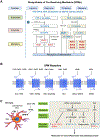
Figure 4:. Schematic for microglial regulation of the resolution of neuroinflammation.
(A) Biosynthesis of SPM families of lipoxins, resolvins, protectins/neuroprotectins, and maresins from DHA and EPA. (B) SPMs receptors. ChemR23 (RvE1 receptor), GPR32 (RvD1 receptor), and GPR18 (RvD2 receptor) are denoted ERV, DRV1, and DRV2, respectively, as with the system of nomenclature for eicosanoid receptors). (C) Microglial signaling via SPM receptors, CB2, and aryl hydrocarbon receptor (AHR), in response to their ligands such as SPMs, cannabinoid, and gut-derived metabolites such as tryptophan (TRP). Activation of these microglial receptors enhances phagocytic activity, reduces the expression of pro-inflammatory mediators (e.g., TNF, IL-1β, and VEGF-B), and increases the production of anti-inflammatory mediators (e.g., IL-10, TGF-α, TGF-β) and SPMs, thus promoting the resolution of neuroinflammation and pathological pain.

References

    1. Abbadie C, Lindia JA, Cumiskey AM, Peterson LB, Mudgett JS, Bayne EK, DeMartino JA, MacIntyre DE, and Forrest MJ (2003). Impaired neuropathic pain responses in mice lacking the chemokine receptor CCR2. Proc Natl Acad Sci U S A 100, 7947–7952. - PMC - PubMed
    1. Ajami B, Samusik N, Wieghofer P, Ho PP, Crotti A, Bjornson Z, Prinz M, Fantl WJ, Nolan GP, and Steinman L (2018). Single-cell mass cytometry reveals distinct populations of brain myeloid cells in mouse neuroinflammation and neurodegeneration models. Nat Neurosci 21, 541–551. - PMC - PubMed
    1. Arita M, Bianchini F, Aliberti J, Sher A, Chiang N, Hong S, Yang R, Petasis NA, and Serhan CN (2005). Stereochemical assignment, anti-inflammatory properties, and receptor for the omega-3 lipid mediator resolvin E1. J Exp Med 201, 713–722. - PMC - PubMed
    1. Asatryan A, and Bazan NG (2017). Molecular mechanisms of signaling via the docosanoid neuroprotectin D1 for cellular homeostasis and neuroprotection. J Biol Chem 292, 12390–12397. - PMC - PubMed
    1. Bang S, Xie YK, Zhang ZJ, Wang Z, Xu ZZ, and Ji RR (2018). GPR37 regulates macrophage phagocytosis and resolution of inflammatory pain. J Clin Invest 128, 3568–3582. - PMC - PubMed

Publication types