. 2019 Feb:127:116-124.
doi: 10.1016/j.yjmcc.2018.12.006. Epub 2018 Dec 17.

Atg2, Atg9 and Atg18 in mitochondrial integrity, cardiac function and healthspan in Drosophila

Affiliations

Atg2, Atg9 and Atg18 in mitochondrial integrity, cardiac function and healthspan in Drosophila

Peng Xu et al. J Mol Cell Cardiol. 2019 Feb.

Abstract

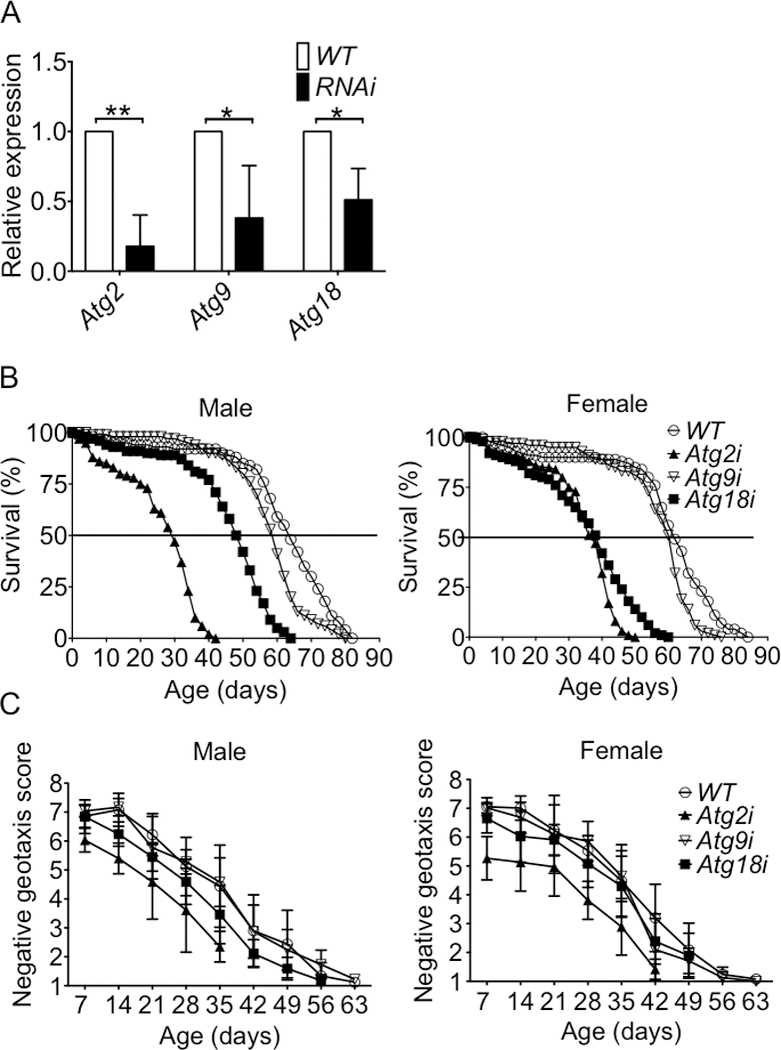
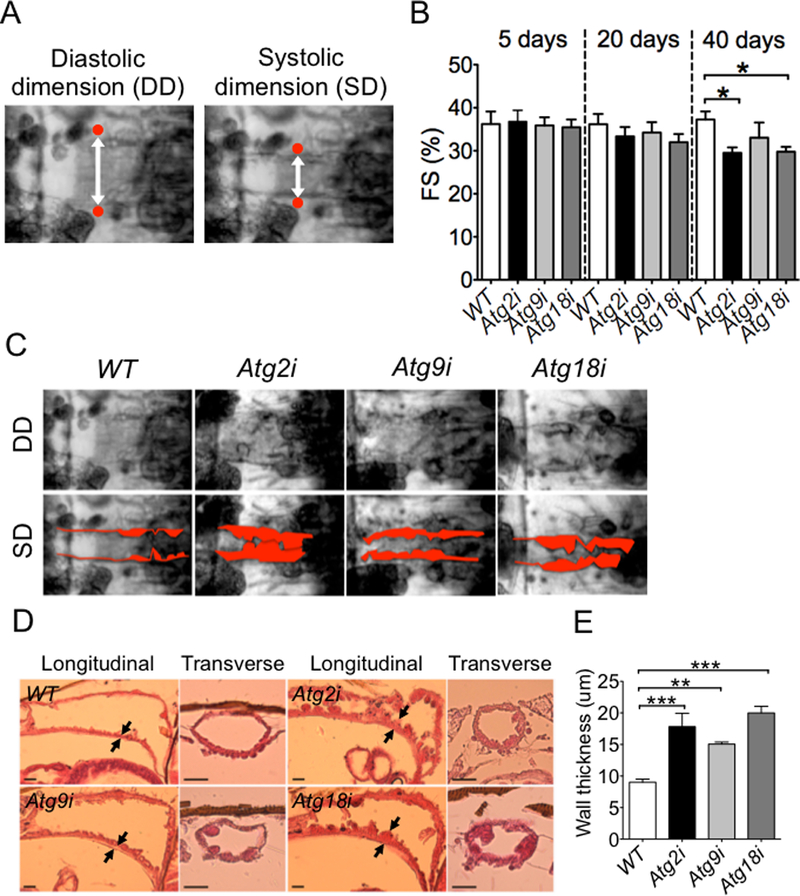
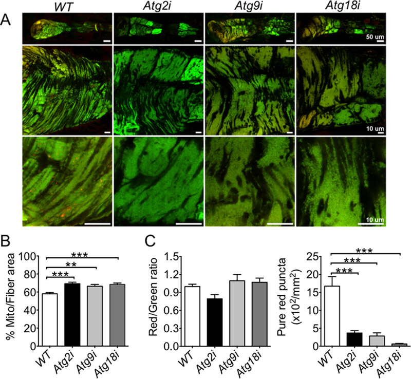
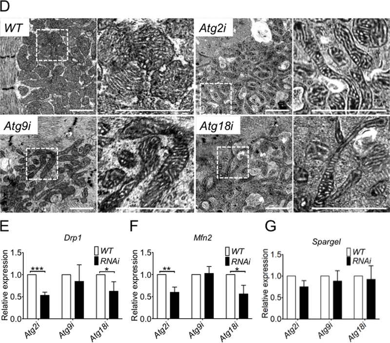
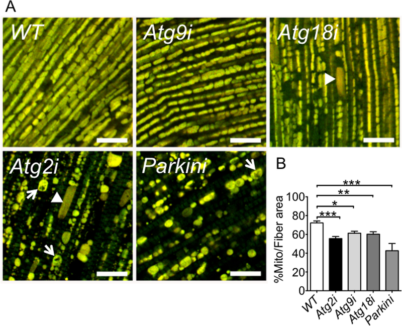
In yeast, the Atg2-Atg18 complex regulates Atg9 recycling from phagophore assembly site during autophagy; their function in higher eukaryotes remains largely unknown. In a targeted screening in Drosophila melanogaster, we show that Mef2-GAL4-RNAi-mediated knockdown of Atg2, Atg9 or Atg18 in the heart and indirect flight muscles led to shortened healthspan (declined locomotive function) and lifespan. These flies displayed an accelerated age-dependent loss of cardiac function along with cardiac hypertrophy (increased heart tube wall thickness) and structural abnormality (distortion of the lumen surface). Using the Mef2-GAL4-MitoTimer mitochondrial reporter system and transmission electron microscopy, we observed significant elongation of mitochondria and reduced number of lysosome-targeted autophagosomes containing mitochondria in the heart tube but exaggerated mitochondrial fragmentation and reduced mitochondrial density in indirect flight muscles. These findings provide the first direct evidence of the importance of Atg2-Atg18/Atg9 autophagy complex in the maintenance of mitochondrial integrity and, regulation of heart and muscle functions in Drosophila, raising the possibility of augmenting Atg2-Atg18/Atg9 activity in promoting mitochondrial health and, muscle and heart function.

Keywords: Autophagy related genes; Cardiac function; Functional aging; Healthspan; Indirect flight muscle; Lifespan; Mitophagy; Negative geotaxis.


Conflict of interest statement

Conflict of Interest

None

Figures

Fig.1.
Fig.1.. Knockdown of Atg2 or Atg18 in both IMF and heart leads to shortened lifespan and reduced locomotor function across the lifespan.
Atg2, Atg9, Atg18 mRNA expression in the heart tube and survival and negative geotaxis were measured in Atg2i, Atg9i and Atg18i flies compared with WT flies across the lifespan. A) One-step SYBR Green-based quantitative RT-PCR analysis of 60 isolated heart tubes. The relative gene expression from an average of three biological replications are normalized to WT (Mef2-Gal4>yw), and significance was determined via an unpaired t-test. B) Survival curve for male and female Atg2i, Atg9i, Atg18i and WT flies; and C) Negative geotaxis score for male and female Atg2i, Atg9i, Atg18i and WT flies. * and ** denote p < 0.05 and p < 0.01, respectively).
Fig. 2.
Fig. 2.. Knockdown of Atg2, Atg9 or Atg18 leads to age-dependent cardiac dysfunction and hypertrophy.
Cardiac function was measured by video-based imaging analysis at 5, 20 and 40 days of age, and heart wall thickness and structure were measure by H&E staining of the heart tube at 40 days of age. A) Captured frames of video images of a fly heart tube at the second abdominal segment. White double head arrows indicate the diastolic and systolic dimensions of the heart tube. Red dots indicate the heart tube edges; B) Quantification of fractional shortening in Atg2i, Atg9i, Atg18i and WT flies (n = 11–15). * denotes p < 0.05; C) Representative light microscope images of 40-day old hearts at the second abdominal segment during a cardiac contraction cycle. Pseudo-red color highlights the heart tube wall; D) H&E stained longitudinal and transverse sections show heart wall thickness. Arrows indicate where the wall thickness was measured. Scale bar = 20 μm; and E) Quantification of the heart wall thickness (n = 4–8). ** and *** denote p < 0.01 and p < 0.001, respectively.
Fig. 3.
Fig. 3.. Knockdown of Atg2, Atg18 or Atg9 leads to reduced mitophagy and mitochondrial abnormalities in cardiomyocytes.
Confocal microscopy and transmission electron microscopy were performed for the heart tubes in Atg knockdown and WT control flies at 40 days of age. A) Representative images of MitoTimer signals (merged image of both GFP and DsRed channels) in cardiomyocyte for assessment of the structure of mitochondrial network and mitochondrial oxidative stress. B) Quantification of mitochondrial volume density (% occupancy of MitoTimer signal) in the A1 heart tube segment (n = 13–20). *** denotes p < 0.001; C) Quantification of Red:Green ratio and number of pure red puncta (n = 13–20). *** denotes p < 0.001; and D) Representative images of transmission electron micrographs. Scale bars = 1 μm; and E, F and G) RT-PCR analysis of Drp1, Mfn2 and Spargel mRNA in Atg2, Atg9, and Atg18 in the heart tubes of 60 knockdown flies (n = 3). *, ** and *** denote p < 0.05, p < 0.01 and p < 0.001, respectively.
Fig. 4.
Fig. 4.. Knockdown of Atg2, Atg9 or Atg18 results in reduced mitochondrial density and abnormal mitochondrial structure in IFM.
Confocal microscopy was performed for IFMs in Atg knockdown and WT control flies at 40 days of age along with Parkin knockdown flies as positive control. A) Representative images of MitoTimer signals (merged image of both GFP and DsRed channels) in IFM. Knockdown of Atg2 show increased numbers of fragmented (arrow head) and hollow mitochondria (arrows), similar to Parkin knockdown flies. Scale bar =10 μm; and B) Quantification of mitochondrial density in IFM (n = 14. *, ** and *** denote p < 0.05, p < 0.01 and p < 0.001, respectively.

References

    1. Klionsky DJ, Emr SD. Autophagy as a regulated pathway of cellular degradation. Science 2000; 290: 1717–1721. - PMC - PubMed
    1. Kim J, Klionsky DJ. Autophagy, cytoplasm-to-vacuole targeting pathway, and pexophagy in yeast and mammalian cells. Annu Rev Biochem 2000; 69: 303–342. - PubMed
    1. Hosokawa N, Hara T, Kaizuka T, Kishi C, Takamura A, Miura Y, et al. Nutrient-dependent mTORC1 association with the ULK1-Atg13-FIP200 complex required for autophagy. Mol Biol Cell 2009; 20: 1981–1991. - PMC - PubMed
    1. Ganley IG, Lam du H, Wang J, Ding X, Chen S, Jiang X. ULK1.ATG13.FIP200 complex mediates mTOR signaling and is essential for autophagy. J Biol Chem 2009; 284: 12297–12305. - PMC - PubMed
    1. Noda T, Suzuki K, Ohsumi Y. Yeast autophagosomes: de novo formation of a membrane structure. Trends Cell Biol 2002; 12: 231–235. - PubMed

Publication types

LinkOut - more resources