Case Reports
. 2018 Dec 19;19(12):4118.
doi: 10.3390/ijms19124118.

Oligoclonal T Cells Transiently Expand and Express Tim-3 and PD-1 Following Anti-CD19 CAR T Cell Therapy: A Case Report

Affiliations
Case Reports

Oligoclonal T Cells Transiently Expand and Express Tim-3 and PD-1 Following Anti-CD19 CAR T Cell Therapy: A Case Report

Christopher Ronald Funk et al. Int J Mol Sci. .

Abstract

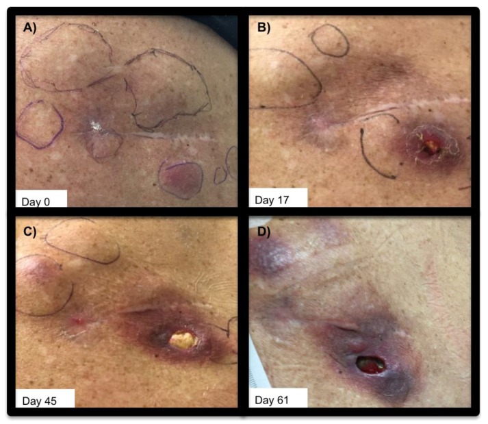
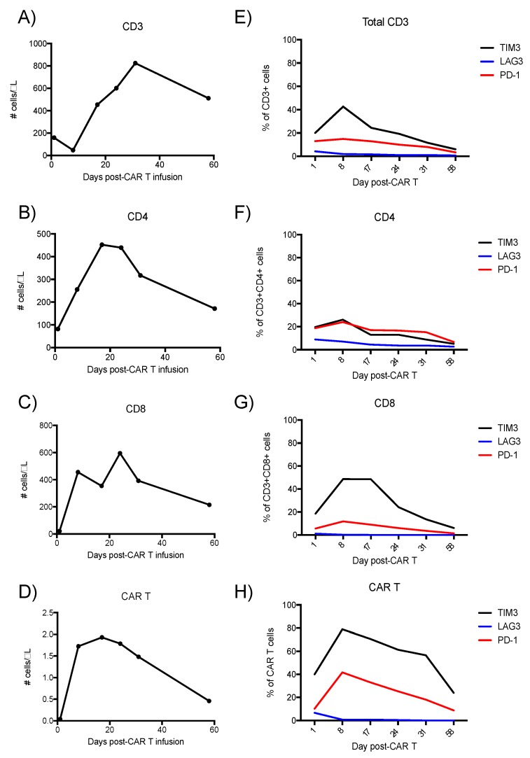
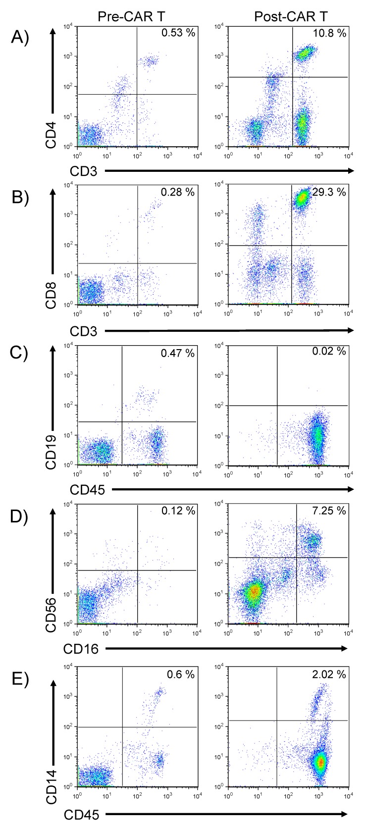
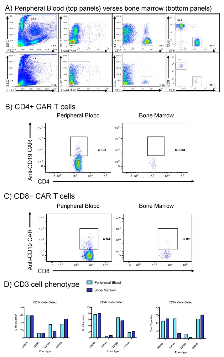
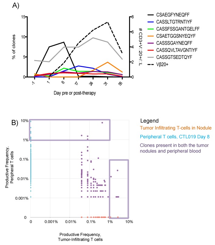
Clinical trials of chimeric antigen receptor (CAR) T cells in hematologic malignancy associate remissions with two profiles of CAR T cell proliferation kinetics, which differ based upon costimulatory domain. Additional T cell intrinsic factors that influence or predict clinical response remain unclear. To address this gap, we report the case of a 68-year-old woman with refractory/relapsed diffuse large B cell lymphoma (DLBCL), treated with tisagenlecleucel (anti-CD19), with a CD137 costimulatory domain (4-1BB) on an investigational new drug application (#16944). For two months post-infusion, the patient experienced dramatic regression of subcutaneous nodules of DLBCL. Unfortunately, her CAR T exhibited kinetics unassociated with remission, and she died of DLBCL-related sequelae. Serial phenotypic analysis of peripheral blood alongside sequencing of the β-peptide variable region of the T cell receptor (TCRβ) revealed distinct waves of oligoclonal T cell expansion with dynamic expression of immune checkpoint molecules. One week prior to CAR T cell contraction, T cell immunoglobulin mucin domain 3 (Tim-3) and programmed cell death protein 1 (PD-1) exhibited peak expressions on both the CD8 T cell (Tim-3 ≈ 50%; PD-1 ≈ 17%) and CAR T cell subsets (Tim-3 ≈ 78%; PD-1 ≈ 40%). These correlative observations draw attention to Tim-3 and PD-1 signaling pathways in context of CAR T cell exhaustion.

Keywords: CTL019; DLBCL; T cell immunoglobulin mucin domain 3 (Tim-3); oligoclonal T cell expansion; programmed cell death protein 1 (PD-1); tisagenlecleucel.


Conflict of interest statement

The authors declare no conflict of interest.

Figures

Figure 1
Figure 1
Subcutaneous DLBCL lesions pre- and post- CAR T cell infusion. Subcutaneous DLBCL lesions superficial to right scapula, shown (A): prior to CAR T infusion (day 0) (B): 17 days post-infusion of CAR T cells, (C): 45 days post-CAR T, and (D): day 61 post-CAR T infusion. Left is medial, and right is lateral.
Figure 2
Figure 2
Transient expansion of T-cells and CAR T cells after tisagenlecleucel CAR T infusion. Panels A–D: Flow cytometry of PBMCs derived from peripheral blood, assessing expression of (A): CD3 (B): CD4 (C): CD8, and (D): CAR. Panels (EH): Expression of immune checkpoint regulators on the T-cells over the same 58 days post-tisagenlecleucel infusion.
Figure 3
Figure 3
Phenotypic analysis of peripheral blood pre- and post-CAR T infusion. Flow cytometry was performed upon peripheral blood to enable phenotypic analysis of immune system cells, including (A): expression of CD3 and CD4 on T cells, (B): expression of CD3 and CD8 on T cells, (C): CD19 and CD45 expression on B-lymphocytes (D): CD56 and CD16 expression on NK cells or cytotoxic T cells, and (E): expression of CD14 and CD45 on monocytes. “Pre-CAR T” denotes analysis performed following lymphodepleting chemotherapy but prior to administration of CAR T cells, whereas “Post-CAR T” denotes analysis on post-CAR T cell infusion day 31.
Figure 4
Figure 4
Comparison of bone marrow aspirates and peripheral blood during episode of pancytopenic aplasia. (A): Whole peripheral blood (top) was compared to bone marrow aspirates (bottom row) with qualitative finding of reduced marrow cellular content. (B,C): CD4+/CD8+ CAR T cells, respectably, were detectable in the peripheral blood and to a minimal extent in the marrow. (D): Comparison of memory/activated T cell (CD45RO+), naïve T cell (CD45RA+), and CD27/28 co-expression in bone marrow (dark blue) and peripheral blood (light blue).
Figure 5
Figure 5
Transient expansion of T cell clones, present at high frequency within the tumor. (A): Deep sequencing of the TCRβ CDR3 regions as performed by Adaptive Biotechnologies (Seattle, WA, USA) on PBMCs collected one day prior to CAR T administration, then on post-infusion days 1, 8, 17, 24, 31, and 58. Deep sequencing data are presented as the productive frequency of clones containing the indicated amino acid sequences in the CDR3 region at each time point. The expansion of CAR T cells was determined by flow cytometry against antibody targeting the Vβ-20+ sequence in the CAR (dashed line). (B): Pair-wise scatter plot showing the productive frequency of the sum of frequencies of clones of T cells, based upon the CDR3 sequencing data. X-axis represents T cells collected from a scapular nodule on post-CAR day 10, while y-axis denotes peripheral blood collected on post-CAR day 8. The violet boxes denote preferential expansion of small numbers of T cell clones, denoted oligoclonal expansion.

References

    1. Porter D.L., Hwang W.-T., Frey N.V., Lacey S.F., Shaw P.A., Loren A.W., Bagg A., Marcucci K.T., Shen A., Gonzalez V., et al. Chimeric antigen receptor T cells persist and induce sustained remissions in relapsed refractory chronic lymphocytic leukemia. Sci. Transl. Med. 2015;7:303ra139. doi: 10.1126/scitranslmed.aac5415. - DOI - PMC - PubMed
    1. Maude S.L., Frey N., Shaw P.A., Aplenc R., Barrett D.M., Bunin N.J., Chew A., Gonzalez V.E., Zheng Z., Lacey S.F., et al. Chimeric Antigen Receptor T Cells for Sustained Remissions in Leukemia. N. Engl. J. Med. 2014;371:1507–1517. doi: 10.1056/NEJMoa1407222. - DOI - PMC - PubMed
    1. Park J.H., Rivière I., Gonen M., Wang X., Sénéchal B., Curran K.J., Sauter C., Wang Y., Santomasso B., Mead E., et al. Long-Term Follow-up of CD19 CAR Therapy in Acute Lymphoblastic Leukemia. N. Engl. J. Med. 2018;378:449–459. doi: 10.1056/NEJMoa1709919. - DOI - PMC - PubMed
    1. Schuster S.J., Bishop M.R., Tam C.S., Waller E.K., Borchmann P., McGuirk J.P., Jaeger U., Jaglowski S., Andreadis C., Westin J.R., et al. Primary Analysis of Juliet: A Global, Pivotal, Phase 2 Trial of CTL019 in Adult Patients with Relapsed or Refractory Diffuse Large B-Cell Lymphoma. Blood. 2017;130:577.
    1. Shimabukuro-Vornhagen A., Gödel P., Subklewe M., Stemmler H.J., Schlößer H.A., Schlaak M., Kochanek M., Böll B., Von Bergwelt-Baildon M.S. Cytokine release syndrome. J. Immunother Cancer. 2018;6:56. doi: 10.1186/s40425-018-0343-9. - DOI - PMC - PubMed

Publication types

Substances