Review
. 2018 Dec 19;7(12):278.
doi: 10.3390/cells7120278.

Autophagy: An Essential Degradation Program for Cellular Homeostasis and Life

Affiliations
Review

Autophagy: An Essential Degradation Program for Cellular Homeostasis and Life

Yoomi Chun et al. Cells. .

Abstract

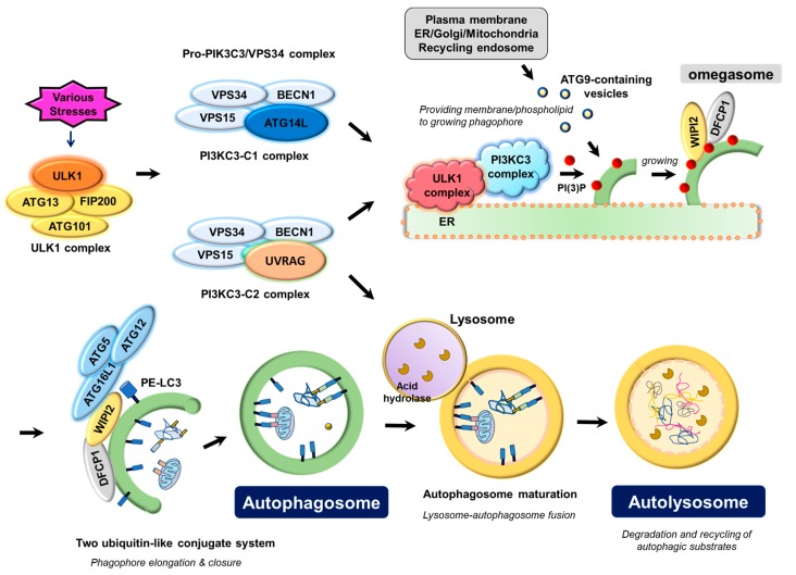
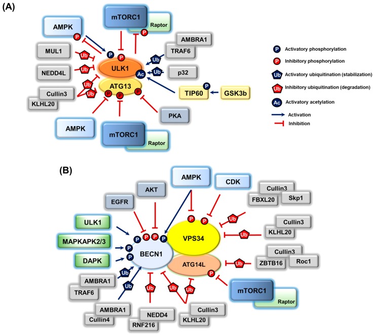
Autophagy is a lysosome-dependent cellular degradation program that responds to a variety of environmental and cellular stresses. It is an evolutionarily well-conserved and essential pathway to maintain cellular homeostasis, therefore, dysfunction of autophagy is closely associated with a wide spectrum of human pathophysiological conditions including cancers and neurodegenerative diseases. The discovery and characterization of the kingdom of autophagy proteins have uncovered the molecular basis of the autophagy process. In addition, recent advances on the various post-translational modifications of autophagy proteins have shed light on the multiple layers of autophagy regulatory mechanisms, and provide novel therapeutic targets for the treatment of the diseases.

Keywords: AMPK-mTORC1-ULK1 triad; PIK3C3/VPS34; autophagy; autophagy adaptor; regulatory modifications.


Conflict of interest statement

The authors declare no conflict of interest.

Figures

Figure 1
Figure 1
Molecular mechanism underlying autophagosome biogenesis by the coordinated actions of ATG proteins. Upon various stresses, the ULK1 complex, consisting of the catalytic subunit ULK1 protein kinase and its associated-regulatory subunits such as ATG13, FIP200, and ATG101, triggers nucleation of the phagophore by phosphorylating and activating the pro-autophagy PIK3C3/VPS34 lipid kinase complex containing either ATG14L (PI3KC3-C1) or UVRAG (PI3KC3-C2), which in turn marks a distinct ER membrane with its phospholipid product, PI(3)P, to form omegasome. PI(3)P on omegasomes then recruits the PI(3)P effector proteins, WIPI2 (WD repeat domain phosphoinositide-interacting protein 2) and DFCP1 (zinc-finger FYVE domain-containing protein 1). WIPI2 and DFCP1 function to gather two ubiquitin-like conjugate complexes, ATG12-ATG5-ATG16L1 and phosphatidylethanolamine (PE)-conjugated LC3 (LC3-II) for elongation and closure of the phagophore membrane. Plasma membrane, mitochondria, recycling endosomes, or Golgi complex may contribute to the elongation of the autophagosomal membrane by providing part of their membrane layers via ATG9. Closure of the phagophore membrane gives rise to a double-membrane bounded vesicle called the autophagosome, which matures and finally fuses with the lysosome to form the autolysosome. Acidic hydrolases in the lysosome degrade the autophagic cargo, and the degradative products are recycled to cope with the stresses that the cells encounter.
Figure 2
Figure 2
A schematic diagram of the selective autophagy for mitochondria, PINK1-Parkin dependent mitophagy. As a central place dictating cell survival and death, the clearance of damaged mitochondria by autophagy (mitophagy) is particularly important. Here, the PINK1-Parkin mediated ubiquitin-dependent pathway is introduced. In response to mitochondrial damage by depolarizing mitochondrial potential, PINK1 is no longer processed by a set of mitochondrial protease systems, and PINK1 is stabilized to accumulate on the mitochondria. And then, PINK1 recruits and activates Parkin by phosphorylating both ubiquitin and Parkin. Activated Parkin on mitochondria poly-ubiquitinates (mostly, Lys63-linked chain) myriad proteins on the damaged mitochondria. Mitophagy adaptors such as OPTN, NDP52, and p62/SQSTM1 function as a bridge between these poly-ubiquitin chains on the damaged mitochondria (via their UBD domain) and LC3 on the autophagosome (via their LIR motif). TBK1 is a protein kinase activated by mitochondrial damage, and phosphorylates the mitophagy adaptor OPTN to increase the binding affinity between OPTN and poly-ubiquitin chains on the dysfunctional mitochondria, thereby, accelerating mitophagy.
Figure 3
Figure 3
Post-translational modifications and regulations of two key autophagy-initiating kinase complexes, the ULK1 and PIK3C3/VPS34 complex. (A) The ULK1 complex is regulated by phosphorylation and is activated by multiple phosphorylations on a catalytic subunit ULK1 by AMPK, and inhibited by phosphorylation on ULK1 (mTORC) as well as ATG13 (mTORC1, PKA, and AMPK). Additionally, the ULK1 complex is activated by ubiquitination on ULK1 (AMBRA1-TRAF6 and p32), whereas it is negatively regulated by ubiquitination-dependent degradation (ULK1 by MUL1, NEDD4L, and Cullin3-KLHL20, ATG13 by Cullin3-KLHL20). In response to growth factor depletion, acetylation of ULK1 is increased by the activated GSK3-TIP60 acetyltransferase axis, resulting in autophagy induction. ULK1 may constitute a negative feedback loop to its upstream regulators, AMPK and mTORC1, by phosphorylating the mTORC1 subunit Raptor protein and all AMPK complex subunits. (B) Phosphoregulation of the PIK3C3/VPS34 complex is observed in the catalytic subunit VPS34 lipid kinase (AMPK and CDK for inhibition), BECN1 (AMPK, ULK1, MAPKAPK2/3, and DAPK for activation; AKT/PKB and EGFR for inhibition), ATG14L/Barkor (mTORC1 for inhibition), and UVRAG (mTORC1 for inhibition). Ubiquitinations on VPS34 (FBXL20-Skp1-Cullin1 and Cullin3-KLHL20 for degradation), BECN1 (NEDD4-RNF216 and Cullin3-KLHL20 for degradation, AMBRA1-TRAF6 or Cullin4 for stabilization), and ATG14L/Barkor (ZBTB16-Cullin3-Roc1 and Cullin3-KLHL20 for degradation) are also important for autophagy regulation.

References

    1. Boya P., Reggiori F., Codogno P. Emerging regulation and functions of autophagy. Nat. Cell Biol. 2013;15:713–720. doi: 10.1038/ncb2788. - DOI - PMC - PubMed
    1. Sridhar S., Botbol Y., Macian F., Cuervo A.M. Autophagy and disease: Always two sides to a problem. J. Pathol. 2012;226:255–273. doi: 10.1002/path.3025. - DOI - PMC - PubMed
    1. Cuervo A.M., Macian F. Autophagy, nutrition and immunology. Mol. Aspects Med. 2012;33:2–13. doi: 10.1016/j.mam.2011.09.001. - DOI - PMC - PubMed
    1. Galluzzi L., Pietrocola F., Levine B., Kroemer G. Metabolic control of autophagy. Cell. 2014;159:1263–1276. doi: 10.1016/j.cell.2014.11.006. - DOI - PMC - PubMed
    1. Catarino S., Pereira P., Girao H. Molecular control of chaperone-mediated autophagy. Essays Biochem. 2017;61:663–674. doi: 10.1042/EBC20170057. - DOI - PubMed

LinkOut - more resources