

Military trainees can accurately measure optic nerve sheath diameter after a brief training session
- PMID: 30572931
- PMCID: PMC6300875
- DOI: 10.1186/s40779-018-0189-y
Military trainees can accurately measure optic nerve sheath diameter after a brief training session
Abstract
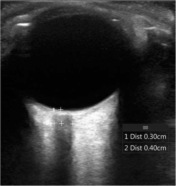
Background: Identification of elevated intracranial pressure is important following traumatic brain injury. We assessed the feasibility of educating military trainees on accurately obtaining optic nerve sheath diameter measurements using a brief didactic and hands-on training session. Optic nerve sheath diameter is a noninvasive surrogate marker for elevated intracranial pressure, and may be of value in remote military operations, where rapid triage decisions must be made without access to advanced medical equipment.
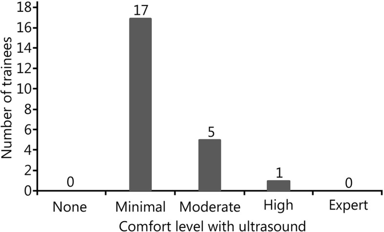
Methods: Military trainees with minimal ultrasound experience were given a 5-min didactic presentation on optic nerve sheath diameter ultrasound. Trainees practiced optic nerve sheath diameter measurements guided by emergency physician ultrasound experts. Trainees then measured the optic nerve sheath diameter on normal volunteers. Following this, a trained physician measured the optic nerve sheath diameter on the same volunteer as a criterion standard. An average of three measurements was taken.
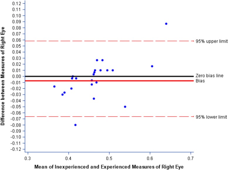
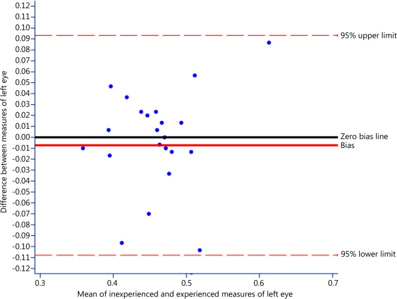
Results: Twenty-three military trainees were enrolled. A mixed design ANOVA was used to compare measurements by trainees to those of physicians, with a mean difference of - 0.6 mm (P = 0.76). A Bland-Altman analysis showed that the degree of bias in optic nerve sheath diameter measures provided by trainees was very small: d = - 0.004 for the right eye and d = - 0.007 for the left eye.
Conclusion: This study demonstrates that optic nerve sheath diameter measurement can be accurately performed by novice ultrasonographers after a brief training session. If validated, point-of-care optic nerve sheath diameter measurement could impact the triage of injured patients in remote areas.
Keywords: Education; Intracranial pressure; Military; Optic nerve sheath diameter; Ultrasound.
Conflict of interest statement
Ethics approval and consent to participate
Not applicable, as all data came from healthy volunteers, and no ED patients were used in this study.
Consent for publication
Not applicable.
Competing interests
The authors declare that they have no competing interests.
Figures
Comment in
-
Ocular ultrasound evaluation of optic nerve sheath diameter in military environments.Mil Med Res. 2019 May 25;6(1):16. doi: 10.1186/s40779-019-0207-8. Mil Med Res. 2019. PMID: 31126318 Free PMC article.
References
MeSH terms
LinkOut - more resources
Full Text Sources
Medical
