. 2019 Mar 15;79(6):1204-1213.
doi: 10.1158/0008-5472.CAN-18-1082. Epub 2018 Dec 20.

Early Noninvasive Detection of Response to Targeted Therapy in Non-Small Cell Lung Cancer

Affiliations

Early Noninvasive Detection of Response to Targeted Therapy in Non-Small Cell Lung Cancer

Jillian Phallen et al. Cancer Res. .

Abstract

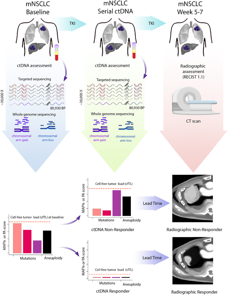
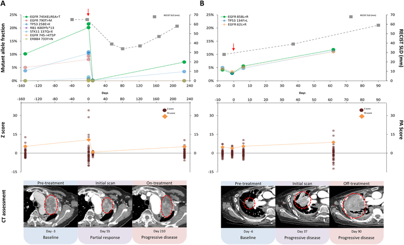
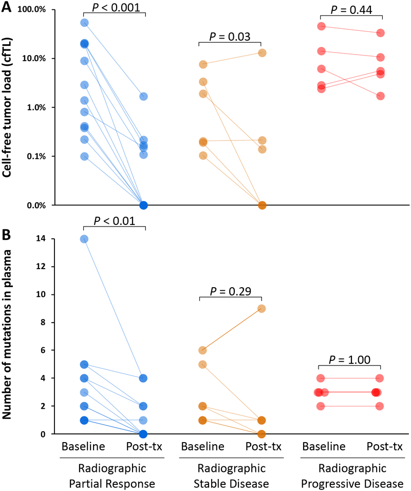
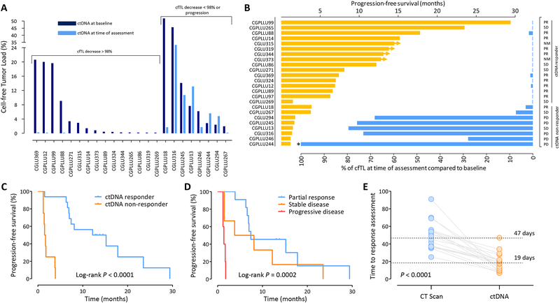
With the advent of precision oncology, there is an urgent need to develop improved methods for rapidly detecting responses to targeted therapies. Here, we have developed an ultrasensitive measure of cell-free tumor load using targeted and whole-genome sequencing approaches to assess responses to tyrosine kinase inhibitors in patients with advanced lung cancer. Analyses of 28 patients treated with anti-EGFR or HER2 therapies revealed a bimodal distribution of cell-free circulating tumor DNA (ctDNA) after therapy initiation, with molecular responders having nearly complete elimination of ctDNA (>98%). Molecular nonresponders displayed limited changes in ctDNA levels posttreatment and experienced significantly shorter progression-free survival (median 1.6 vs. 13.7 months, P < 0.0001; HR = 66.6; 95% confidence interval, 13.0-341.7), which was detected on average 4 weeks earlier than CT imaging. ctDNA analyses of patients with radiographic stable or nonmeasurable disease improved prediction of clinical outcome compared with CT imaging. These analyses provide a rapid approach for evaluating therapeutic response to targeted therapies and have important implications for the management of patients with cancer and the development of new therapeutics.Significance: Cell-free tumor load provides a novel approach for evaluating longitudinal changes in ctDNA during systemic treatment with tyrosine kinase inhibitors and serves an unmet clinical need for real-time, noninvasive detection of tumor response to targeted therapies before radiographic assessment.See related commentary by Zou and Meyerson, p. 1038.


Conflict of interest statement

Disclosure of Potential Conflicts of Interest

P.M.F. has been a consultant for AstraZeneca/Medimmune and Boehringer-Ingelheim. V. Adleff is a consultant for Personal Genome Diagnostics. V.E.V. is a founder of, holds equity in, and is a member of the Board of Directors of Personal Genome Diagnostics (PGDx). Under a license agreement between PGDx and the Johns Hopkins University, V.E.V. is entitled to a share of royalty received by the University on sales of services or products by PGDx. V.E.V. is a member of the Scientific Advisory Board of Ignyta. The terms of these arrangements have been reviewed and approved by the Johns Hopkins University in accordance with its conflict of interest policies. H.H. has served on advisory boards for Astrazeneca, Merck, Boehringer Ingelheim, Abbvie, and Foundation Medicine, speakers honorarium from Bristol Myers Squibb, Merck, and Astrazeneca, and research funding from Pfizer.

Figures

Figure 1.
Figure 1.. Schematic of cfTL determination and prediction of therapeutic response.
Liquid biopsies from metastatic non-small-cell lung cancer (mNSCLC) patients undergoing treatment with tyrosine kinase inhibition (TKI) were analyzed at baseline and at serial time points after treatment. The TEC-Seq approach was used to directly identify sequence alterations across 58 genes encompassing 80,930 bases sequenced to >30,000X coverage, and whole-genome approaches were used to identify copy number changes in cfDNA. Cell-free tumor load (cfTL) was determined as the mutant allele fraction of the most abundant alteration in a clone targeted by TKI for patients with detected sequence alterations, or as the presence or absence of aneuploidy based on PA score in patients without detectable sequence alterations. Prediction of therapeutic response to targeted therapy based on ctDNA dynamics was assessed through changes in cfTL from baseline to subsequent time points treatment whereas response assessment through CT imaging was performed 5–7 weeks after treatment.
Figure 2.
Figure 2.. Dynamic changes of ctDNA during therapy.
Characteristic patterns of ctDNA changes during therapy are shown for a responder (CGPLLU12) (A) and a non-responder (CGPLLU244) (B), both treated with osimertinib. Mutant allele fractions of clones identified in cfDNA through the TEC-Seq approach are shown for each timepoint analyzed with the ctDNA clone representing cfTL shown in bright green and treatment initiation highlighted with a red arrow (Top). Copy number changes identified in cfDNA from analyses of whole-genome data are shown at each timepoint analyzed as Z scores (burgundy dots) for each chromosome arm and PA scores (orange diamonds) (Middle). RECIST 1.1 sum of longest diameters (SLD, gray boxes) were measured from CT scans at intervals during therapy (Top) and CT images show representative tumor lesions for each patient circled in red (Bottom).
Figure 3.
Figure 3.. Characteristics of cell-free DNA in patients treated with tyrosine kinase inhibitors.
Changes in cfTL (A) and the number of mutations in plasma (B) in patients with partial response (blue), stable disease (orange), and progressive disease (red) from baseline to the time of ctDNA assessment after initiation of therapy. The partial response subgroup also included two patients with non-measureable disease by RECIST who were classified as clinical responders.
Figure 4.
Figure 4.. Detection of ctDNA variants within hours after tyrosine kinase inhibitor initiation.
Changes in the levels of ctDNA (A) as well as of cfDNA extracted (B) are depicted for eight patients at baseline and at four to twelve hours after the initiation of targeted therapy. Emerging ctDNA alterations and the corresponding cfDNA amounts for patients with these alterations are depicted in red.
Figure 5.
Figure 5.. Changes in ctDNA and prediction of response to therapy.
Changes in cfTL from baseline to the time of ctDNA assessment revealed a bimodal distribution (A). Patients with reduction of cfTL >98% and ≤98% were categorized as ctDNA responders and ctDNA non-responders, respectively. cfTL at the time of ctDNA assessment (blue) and PFS (orange) are depicted for patients analyzed (B). Radiographic assessment is indicated in the right column as partial response (PR), stable disease (SD), non-measurable disease (NM), or progressive disease (PD). Patient CGPLLU244 had cfTL levels >100% at the time of ctDNA assessment (*). Progression-free survival for ctDNA responders and non-responders (P < 0.0001, Mantel-Cox log rank test) (C). Progression-free survival based on initial radiographic assessment (P = 0.0002, Mantel-Cox log rank test) (D) Time to response assessment as determined by CT scans (blue) or analyses of ctDNA (orange) are indicated with median time to assessment shown in dotted lines (P < 0.0001, Wilcoxon signed rank test) (E).

Comment in

References

    1. Sawyers C Targeted cancer therapy. Nature 2004;432:294–7 - PubMed
    1. Vogelstein B, Papadopoulos N, Velculescu VE, Zhou S, Diaz LA Jr., Kinzler KW. Cancer genome landscapes. Science 2013;339:1546–58 - PMC - PubMed
    1. Haber DA, Velculescu VE. Blood-based analyses of cancer: circulating tumor cells and circulating tumor DNA. Cancer discovery 2014;4:650–61 - PMC - PubMed
    1. Newman AM, Bratman SV, To J, Wynne JF, Eclov NC, Modlin LA, et al. An ultrasensitive method for quantitating circulating tumor DNA with broad patient coverage. Nat Med 2014;20:548–54 - PMC - PubMed
    1. Bettegowda C, Sausen M, Leary RJ, Kinde I, Wang Y, Agrawal N, et al. Detection of circulating tumor DNA in early- and late-stage human malignancies. Sci Transl Med 2014;6:224ra24 - PMC - PubMed

Publication types

MeSH terms