. 2019 Jan;68(1):3-14.
doi: 10.2337/dbi18-0035.

Adipose Tissue, Inter-Organ Communication, and the Path to Type 2 Diabetes: The 2016 Banting Medal for Scientific Achievement Lecture

Affiliations

Adipose Tissue, Inter-Organ Communication, and the Path to Type 2 Diabetes: The 2016 Banting Medal for Scientific Achievement Lecture

Barbara B Kahn. Diabetes. 2019 Jan.

Abstract

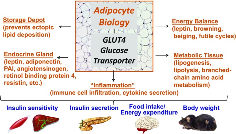
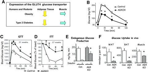
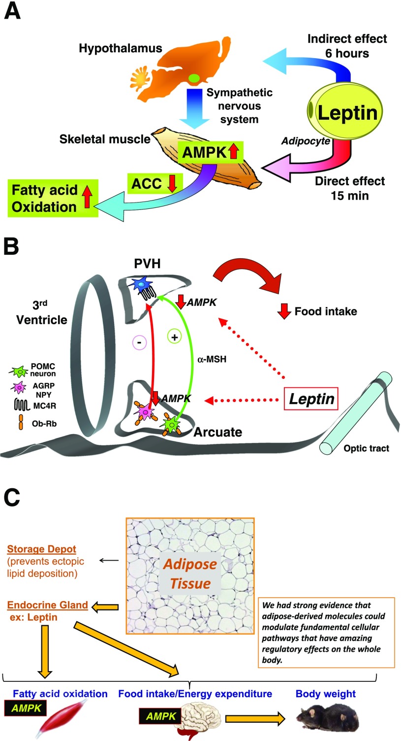
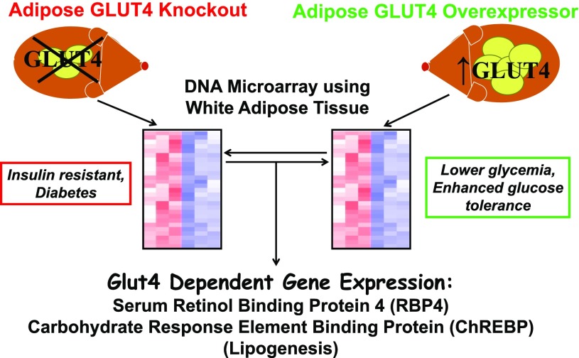
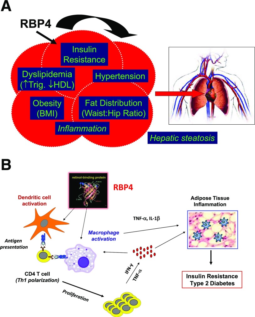
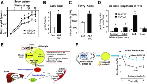
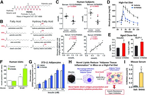
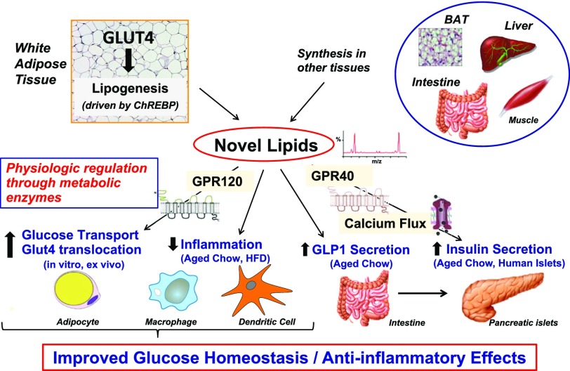
My scientific career has focused on understanding the mechanisms underlying insulin resistance with the goal of developing new strategies to prevent and treat type 2 diabetes. My early studies focused on understanding how insulin promotes glucose transport into adipocytes, a classic model of highly insulin-responsive target cells. When we found changes in adipocyte glucose transport in altered metabolic states, we were highly motivated to understand the consequences of this on whole-body glucose homeostasis. In the late 1980s, when GLUT4, the major insulin-regulated glucose transporter, was identified, my lab observed that it was downregulated in adipocytes but not in skeletal muscle in insulin-resistant states, such as obesity and type 2 diabetes, in humans and rodents. We investigated the role of GLUT4 in adipose tissue and muscle in whole-body insulin sensitivity, making tissue-specific GLUT4-overexpressing and GLUT4 knockout mice. These studies led to the discovery that adipocytes, and specifically glucose transport into adipocytes, regulate whole-body glucose homeostasis. As adipocytes take up relatively little glucose, we investigated the underlying mechanisms. In the 1990s, we performed DNA microarrays on adipose tissue from adipose-specific GLUT4-overexpressing and GLUT4 knockout mice to find reciprocally regulated genes, and we identified several molecules that were not previously known to regulate systemic insulin sensitivity and/or energy balance. More recently, with Alan Saghatelian's lab, we discovered a novel class of lipids with antidiabetes and anti-inflammatory effects. We also investigated the effects of the adipose-secreted hormone, leptin, on insulin sensitivity. We found that the AMP-activated protein kinase (AMPK) pathway mediates leptin's effects on fatty acid oxidation in muscle and also plays a role in leptin's anorexigenic effects in the hypothalamus. These studies transformed AMPK from a "fuel gauge" that regulates energy supply at the cellular level to a sensing and signaling pathway that regulates organismal energy balance. Overall, these studies have expanded our understanding of the multifaceted role of adipose tissue in metabolic health and how adipose dysfunction increases the risk for type 2 diabetes.


Figures

Figure 1
Figure 1
White adipose tissue regulates whole-body metabolism. The adipocyte is not only a storage depot but has many other functions including 1) as an endocrine gland, secreting many hormones, cytokines, and vasoactive substances; 2) having a role in energy balance, by secreting hormones such as leptin and also by browning and beiging and the futile cycles that burn energy; and 3) as a metabolic tissue that performs lipogenesis, lipolysis, and metabolism of branched-chain amino acids and other molecules. Adipose inflammation, characterized by immune cell infiltration and cytokine secretion, occurs in the setting of obesity and insulin resistance. All of these processes contribute to regulation of insulin sensitivity, insulin secretion, food intake and energy expenditure, and ultimately body weight. PAI, plasminogen activator inhibitor.
Figure 2
Figure 2
Role of GLUT4 in regulating whole-body glucose tolerance and insulin sensitivity. GLUT4 protein levels are decreased in white adipose tissue but not in skeletal muscle, of insulin-resistant humans and rodents (A). Adipose-specific overexpression of GLUT4 (AG4OX) lowers fasting glycemia and improves whole-body glucose tolerance (B), whereas adipose-specific GLUT4 deletion (AG4KO) causes whole-body glucose intolerance (C) and insulin resistance (DF). Adipose-specific GLUT4 deletion causes liver insulin resistance (E) and decreases glucose uptake in adipose tissue and skeletal muscle (F). For experimental details for panel B, see Carvalho et al. (4). For experimental details for panels CF, see Abel et al. (5). Panel B reprinted from Carvalho et al. (4). Panels CF reprinted from Abel et al. (5). *Panels BD and F: P < 0.05 vs. control; panel E: P < 0.05 vs. control + insulin. BAT, brown adipose tissue; Con, control; GTT, glucose tolerance test; ITT, insulin tolerance test; WAT, white adipose tissue.
Figure 3
Figure 3
Leptin regulates fatty acid oxidation in muscle by activating AMPK. A: Leptin activates α2-AMPK in red skeletal muscle through two distinct mechanisms. Early activation of AMPK after intravenous leptin administration appears to be direct, whereas later activation depends on the hypothalamic–sympathetic nervous system axis. Leptin’s effect to activate AMPK is associated with suppression of acetyl-CoA carboxylase (ACC) activity and results in stimulation of fatty acid oxidation. Panel A reprinted from Minokoshi et al. (10). B: Leptin inhibits α2-AMPK activity in the arcuate and paraventricular hypothalamus (PVH), and this is necessary to decrease food intake in response to leptin. Panel B reprinted from Minokoshi et al. (11). C: Schematic of how white adipose tissue regulates whole-body metabolism. Through its endocrine actions—in this case, leptin—white adipose tissue can modulate AMPK signaling in other tissues. This is fundamental to maintaining energy supplies at the cellular level, and this pathway stimulates fatty acid oxidation in muscle. In addition, leptin secreted from white adipose tissue inhibits the AMPK pathway in the hypothalamus to regulate food intake and energy expenditure and ultimately body weight. Our early work with leptin and AMPK provided strong evidence that adipose-derived molecules could modulate fundamental cellular pathways that have regulatory effects on the whole body.
Figure 4
Figure 4
Microarray analysis of adipose tissue from adipose-specific GLUT4 knockout and adipose-specific GLUT4 overexpressor mice. White adipose tissue from these genetically modified mice was subjected to Affymetrix DNA array analysis. This led to the identification of GLUT4-dependent genes, which have systemic effects on insulin sensitivity. Examples are RBP4, the major protein transporting retinol in the blood, and ChREBP, a transcription factor that regulates lipogenesis and glycolysis.
Figure 5
Figure 5
Role of RBP4 in regulating whole-body insulin sensitivity. A: Serum RBP4 levels correlate with multiple components of the metabolic syndrome. B: RBP4-induced activation of antigen-presenting cells in adipose tissue is sufficient to cause adipose “inflammation” and systemic insulin resistance. Panel B reprinted from Moraes-Vieira et al. (25). Trig., triglycerides.
Figure 6
Figure 6
Role of GLUT4 in the regulation of adiposity and lipid metabolism. Adipose-specific overexpression of GLUT4 (AG4OX) results in increased body weight (A), adiposity (B), serum free fatty acids (C), and de novo lipogenesis in white and brown adipose depots but not in liver (D). Panels A and B reprinted from Shepherd et al. (3). Panel C adapted from Table 2 in Carvalho et al. (4). In panel D, some data are adapted from Herman et al. (28). E: Increasing glucose transport in adipocytes enhances systemic insulin sensitivity by channeling glucose into fatty acid synthesis. When GLUT4 protein levels increase, ChREBP senses the increased glucose transport, leading to alterations in adipocyte biology, including driving more glucose into fatty acid synthesis (also known as de novo lipogenesis). That process has favorable effects on insulin action to suppress hepatic glucose production and on insulin signaling and glucose utilization in muscle. Knocking out (KO) ChREBP reverses the increased fatty acid synthesis and the beneficial effects of increased GLUT4 expression in adipocytes (see box insert). M.A. Herman contributed to the original design of panel E. F: Adipose-specific KO of ChREBP in otherwise normal mice reduces fatty acid synthesis and causes systemic insulin resistance. Data in panel F was reprinted from Vijayakumar et al. (29). For experimental details for panels A and B, see Shepherd et al. (3); for panel C, see Carvalho et al. (4); for panel D, see Herman et al. (28); and for panel F, see Vijayakumar et al. (29). BAT, brown adipose tissue; Con, control; SubQ, subcutaneous adipose tissue. *Panels AD and F: P < 0.05 vs. control.
Figure 7
Figure 7
FAHFAs are a novel class of lipids with beneficial metabolic and anti-inflammatory effects. A: FAHFAs consist of a fatty acid group, such as palmitic acid, and a hydroxy fatty acid group joined by an ester bond (PAHSA). B: FAHFAs can consist of multiple different fatty acids and their hydroxy fatty acids in various combinations. Examples of some constituent fatty acids are shown. C: PAHSA levels in serum are lower in insulin-resistant people than in the insulin-sensitive people and serum levels correlate highly with insulin sensitivity determined by glucose infusion rate during a euglycemic-hyperinsulinemic clamp. A single oral dose of PAHSA improves glucose tolerance of high-fat diet–fed mice (D) and augments insulin and GLP1 secretion in aged, insulin-resistant chow-fed mice (E). These secretory effects are not seen in high-fat diet–fed mice. PAHSAs enhance glucose-stimulated insulin secretion directly in human islets (F) and enhance insulin-stimulated glucose transport in cultured adipocytes (G). H: Anti-inflammatory effects of PAHSAs include blocking antigen presentation, proinflammatory cytokine production, and costimulatory molecule production. This leads to decreased adipose tissue inflammation and insulin resistance. I: Administration of 9HHA, a nonnatural hydroxy fatty acid, results in the synthesis of 9PAHHA, proving that PAHSA can be synthesized in vivo. For experimental details for panels AG and I, see Yore et al. (30). Panels AG and I reprinted from Yore et al. (30). *Panel C: P < 0.0005 vs. insulin-sensitive subjects; panels D, E, and I: P < 0.05 vs. vehicle (Veh); panel F: P < 0.05 vs. 2.5 mmol/L glucose, same treatment; panel G: P < 0.05 vs. DMSO, same insulin concentration. #P < 0.05 vs. control, same glucose concentration. DPM, disintegrations per minute; LBM, lean body mass.
Figure 8
Figure 8
Summary of biologic activities of FAHFAs. Increased GLUT4 protein levels in adipocytes results in increased de novo lipogenesis driven by ChREBP. This results in the production of FAHFAs, which act through GPCRs (GPR-120) to augment glucose transport and GLUT4 translocation. These lipids also decrease inflammatory responses in macrophages and dendritic cells. In aged, insulin-resistant chow-fed mice (but not in high-fat diet [HFD]–fed mice), these lipids increase glucose-stimulated GLP1 secretion and insulin secretion. FAHFAs directly increase insulin secretion in human pancreatic β-cells, which involves GPR-40. As we do not see these secretory effects of the novel lipids in mice on an HFD but find increased insulin sensitivity with chronic PAHSA treatment, it is likely that insulin-sensitizing effects of the PAHSAs play a major role in their beneficial effects in HFD-fed mice. Adapted from Yore et al. (30). BAT, brown adipose tissue.

Similar articles

Cited by

References

    1. Herman MA, She P, Peroni OD, Lynch CJ, Kahn BB. Adipose tissue branched chain amino acid (BCAA) metabolism modulates circulating BCAA levels. J Biol Chem 2010;285:11348–11356 - PMC - PubMed
    1. Ross SR, Graves RA, Spiegelman BM. Targeted expression of a toxin gene to adipose tissue: transgenic mice resistant to obesity. Genes Dev 1993;7:1318–1324 - PubMed
    1. Shepherd PR, Gnudi L, Tozzo E, Yang H, Leach F, Kahn BB. Adipose cell hyperplasia and enhanced glucose disposal in transgenic mice overexpressing GLUT4 selectively in adipose tissue. J Biol Chem 1993;268:22243–22246 - PubMed
    1. Carvalho E, Kotani K, Peroni OD, Kahn BB. Adipose-specific overexpression of GLUT4 reverses insulin resistance and diabetes in mice lacking GLUT4 selectively in muscle. Am J Physiol Endocrinol Metab 2005;289:E551–E561 - PubMed
    1. Abel ED, Peroni O, Kim JK, et al. . Adipose-selective targeting of the GLUT4 gene impairs insulin action in muscle and liver. Nature 2001;409:729–733 - PubMed

Publication types