. 2019 Feb 15;294(7):2386-2396.
doi: 10.1074/jbc.RA118.006226. Epub 2018 Dec 20.

Interleukin 34 (IL-34) cell-surface localization regulated by the molecular chaperone 78-kDa glucose-regulated protein facilitates the differentiation of monocytic cells

Affiliations

Interleukin 34 (IL-34) cell-surface localization regulated by the molecular chaperone 78-kDa glucose-regulated protein facilitates the differentiation of monocytic cells

Sayaka Ogawa et al. J Biol Chem. .

Abstract

Interleukin 34 (IL-34) constitutes a cytokine that shares a common receptor, colony-stimulating factor-1 receptor (CSF-1R), with CSF-1. We recently identified a novel type of monocytic cell termed follicular dendritic cell-induced monocytic cells (FDMCs), whose differentiation depended on CSF-1R signaling through the IL-34 produced from a follicular dendritic cell line, FL-Y. Here, we report the functional mechanisms of the IL-34-mediated CSF-1R signaling underlying FDMC differentiation. CRIPSR/Cas9-mediated knockout of the Il34 gene confirmed that the ability of FL-Y cells to induce FDMCs completely depends on the IL-34 expressed by FL-Y cells. Transwell culture experiments revealed that FDMC differentiation requires a signal from a membrane-anchored form of IL-34 on the FL-Y cell surface, but not from a secreted form, in a direct interaction between FDMC precursor cells and FL-Y cells. Furthermore, flow cytometric analysis using an anti-IL-34 antibody indicated that IL-34 was also expressed on the FL-Y cell surface. Thus, we explored proteins interacting with IL-34 in FL-Y cells. Mass spectrometry analysis and pulldown assay identified that IL-34 was associated with the molecular chaperone 78-kDa glucose-regulated protein (GRP78) in the plasma membrane fraction of FL-Y cells. Consistent with this finding, GRP78-heterozygous FL-Y cells expressed a lower level of IL-34 protein on their cell surface and exhibited a reduced competency to induce FDMC differentiation compared with the original FL-Y cells. These results indicated a novel GRP78-dependent localization and specific function of IL-34 in FL-Y cells related to monocytic cell differentiation.

Keywords: CRISPR/Cas; GRP78; Western blot; cell differentiation; cell surface protein; chaperone; colony-stimulating factor-1 receptor; cytokine; follicular dendritic cell; interleukin; interleukin 34; monocyte; plasma membrane.


Conflict of interest statement

The authors declare that they have no conflicts of interest with the contents of this article

Figures

Figure 1.
Figure 1.
Role of IL-34 in FDMC differentiation. A, gene targeting strategy for establishing IL-34 KO FL-Y cells. Exon 3 of the Il34 gene was replaced with the puromycin-resistance marker gene (puror) and the blasticidin S–resistance gene (BSR). Open arrowheads show primer pairs used for detecting successful recombination. B, genomic PCR analysis of the Il34 allele in FL-Y cells. Genome DNA was prepared from IL-34 KO FL-Y cells and analyzed by PCR. Il34 KO alleles were identified using indicated primer pairs (F/puro or F/BSR2). Closed arrowheads on the right show PCR products with the expected sizes. C, RT-PCR analysis of Il34 mRNA expression in IL-34 KO FL-Y cells. mRNA was isolated from WT FL-Y and IL-34 KO FL-Y cells (clones 1–3), and Il34 transcript was amplified by RT-PCR. Alternatively, an exon 3-specific primer (ex3F) was also used to amplify the Il34 transcripts. Closed arrowheads on the right show a PCR product with the expected size. The open arrowhead shows smaller PCR products than the expected one. D, T cell– and adherent cell–depleted splenocytes (1 × 10⁶) from BALB/c mice were cultured with 1 × 10⁴ cells of FL-Y or IL-34 KO FL-Y or without FL-Y (−) for 10 days. Cultured cells were collected and analyzed by flow cytometry after staining for CD11b. The percentage of CD11b+ cells is indicated. E, the number of generated FDMCs in each culture was calculated from the percentage of CD11b+ cells in the total number of viable cells in D. F, T cell– and adherent cell–depleted splenocytes (1 × 10⁶) from BALB/c mice were cultured with 1 × 10⁴ cells of FL-Y in the presence or absence of GW2580 (1 μm). The number of CD11b+ cells was determined as shown in D and E. The data are presented as the means ± S.D. of triplicate cultures. The data are representative of at least three independent experiments. Statistical differences are marked: **, p < 0.01 versus FL-Y; ***, p < 0.005 versus the DMSO control.
Figure 2.
Figure 2.
Involvement of cell-surface IL-34 in FDMC differentiation. A, in the Transwell culture, 1 × 10⁴ FL-Y cells were seeded to the lower chamber, and T cell– and adherent cell–depleted splenocytes (1 × 10⁶ cells) from BALB/c mice were added to the upper chamber separated by a 0.45-μm-pore size membrane. After 9 days, the number of CD11b+ cells in the upper chamber was estimated by flow cytometry. The data are presented as the means ± S.D. of triplicate cultures. Statistical differences are marked: *, p < 0.05 versus intact culture condition for FDMC induction of FL-Y cells. B, T cell– and adherent cell–depleted splenocytes (1 × 10⁶) from BALB/c mice were cultured with 2 × 10⁵ cells of the original FL-Y (FL-Y) or IL-34 KO FL-Y (IL-34 KO) cells, each of which was pretreated with (+) or without (−) 0.1% PFA. After 10 days, the cultured cells were analyzed by flow cytometry after staining for CD11b. The data are presented as the means ± S.D. of triplicate cultures. Statistical differences are marked: **, p < 0.01 versus the original FL-Y treated with PFA. C, IL-34 expression on the cell surface of the FL-Y cell line was determined by flow cytometry (left panels) after staining with isotype IgG (shaded) or anti–IL-34 antibody (red line). The level of IL-34 surface expression is indicated as the ΔMFI value, which was calculated by subtracting the MFI value of the isotype-matched control from that of each sample (right panel). The data are representative of at least three independent experiments. Max, maximum.
Figure 3.
Figure 3.
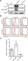
Establishment of FL-Y cells expressing Strep-tagged IL-34. A, immunoblot analysis of IL-34 in FL-Y, FL-Y–IL-34–Nst (IL-34-Nst), and IL-34 KO FL-Y (IL-34 KO) cells. Whole cell lysates were subjected to Western blotting analysis using anti–IL-34, anti–Strep-tag, and anti–β-actin Abs. β-Actin was used as an internal control. The blots are representative of at least three independent experiments. Arrows indicate IL-34–Nst. B, flow cytometric analysis of FL-Y cells after staining with anti–IL-34 or anti–Strep-tag Abs (red line). Negative controls that were stained with an isotype-matched control Ab are shown in gray histograms. C, T cell– and adherent cell–depleted splenocytes (1 × 10⁶) from BALB/c mice were cultured with 2 × 10⁵ cells of WT FL-Y, FL-Y–IL-34–Nst (IL-34-Nst), and IL-34 KO FL-Y (IL-34 KO) cells that were pretreated with 0.1% paraformaldehyde. After 8 days, the cultured cells were analyzed by flow cytometry after staining for CD11b. The data are representative of at least three independent experiments. The data are presented as the means ± S.D. of triplicate cultures. Statistical differences are marked: **, p < 0.01 versus FL-Y; *, p < 0.05 versus FL-Y.
Figure 4.
Figure 4.
Interaction of IL-34 with GRP78 in the plasma membrane of FL-Y cells. A, immunoblot analysis of the plasma membrane fraction prepared from FL-Y–IL-34–Nst (IL-34-Nst) and IL-34 KO FL-Y (IL-34 KO) cells. Plasma membrane fractions extracted from the indicated cells were subjected to Western blotting analysis using anti–IL-34 and anti–Strep-tag Abs. VCAM was used as an internal control. Arrows indicate IL-34–Nst. B, silver staining of IL-34 and proteins associated with IL-34. IL-34–Nst proteins in the plasma membrane fraction prepared from FL-Y–IL-34 (IL-34) and FL-Y–IL-34–Nst (IL-34-Nst) cells were pulled down by using the Strep-Tactin Sepharose resin and eluted by 2.5 mm desthiobiotin. Plasma membrane fractions (Input), eluted fractions with desthiobiotin (EF), and residual fractions on the resin (RF) were subjected to SDS-PAGE. C, GRP78 was bound to IL-34–Nst in the plasma membrane fraction. The plasma membrane fraction of FL-Y–IL-34–Nst cells was mixed with control (Cont) or Strep-Tactin (ST) Sepharose, and bound molecules were eluted with SDS-PAGE sample buffer for Western blotting with anti-GRP78 Ab. D, plasma membrane fraction of FL-Y–IL-34 (IL-34) and FL-Y–IL-34–Nst (IL-34-Nst) cells was reacted with Strep-Tactin Sepharose, and binding molecules were eluted with SDS-PAGE sample buffer for Western blotting with anti-GRP78 Ab. E, recombinant GST or GST–GRP78 was mixed with the whole cell lysate of FL-Y–IL-34–Nst cells and precipitated with glutathione-Sepharose. Eluted fractions were subjected to SDS-PAGE and Western blotting analysis.
Figure 5.
Figure 5.
GRP78-heterozygous FL-Y cells exhibit reduced FDMC-inducing activity. A, gene targeting strategy for establishing GRP78-heterozygous FL-Y cells. Open arrowheads show primer pairs used for detecting successful recombination. B, genomic PCR analysis of the Grp78 gene in WT (+/+) and GRP78-heterozygous (+/−) FL-Y cells. Genome DNA was prepared from GRP78-heterozygous FL-Y cells (clones 1 and 2) and amplified by PCR. The Grp78 KO allele was identified using indicated primer pairs (F/puro and r-plox/R). C, RT-PCR analysis of Grp78 mRNA expression in GRP78-heterozygous FL-Y cells (clones 1 and 2). RNA was isolated from WT and GRP78-heterozygous FL-Y cells (clones 1 and 2), and the Grp78 transcript was amplified by RT-PCR. D, Western blotting analysis of GRP78 expressed in FL-Y and GRP78-heterozygous FL-Y cells (clones 1 and 2). Whole cell lysates prepared from FL-Y and GRP78-heterozygous FL-Y cells (clones 1 and 2) were separated by SDS-PAGE and subjected to Western blotting. E, IL-34 expression on the cell surface of the GRP78-heterozygous FL-Y cell line was determined by flow cytometry after staining with an anti–IL-34 antibody as shown in Fig. 2C. The level of IL-34 cell-surface expression is indicated as described for Fig. 2C (ΔMFI). F, competency of GRP78-heterozygous FL-Y cells for inducing FDMC differentiation. T cell– and adherent cell–depleted splenocytes (1 × 10⁶) from BALB/c mice were cultured with FL-Y and GRP78-heterozygous (±) FL-Y (clones 1 and 2) that were pretreated with 0.1% paraformaldehyde as shown in Fig. 2B. Cultured cells were collected and analyzed by flow cytometry after staining for CD11b. The data are presented as the means ± S.D. of triplicate cultures. The data are representative of at least three independent experiments. Statistical differences are marked: *, p < 0.05 versus original FL-Y (+/+).

Similar articles

Cited by

References

    1. Hume D. A. (2008) Macrophages as APC and the dendritic cell myth. J. Immunol. 181, 5829–5835 10.4049/jimmunol.181.9.5829 - DOI - PubMed
    1. Wei S., Nandi S., Chitu V., Yeung Y.-G., Yu W., Huang M., Williams L. T., Lin H., and Stanley E. R. (2010) Functional overlap but differential expression of CSF-1 and IL-34 in their CSF-1 receptor-mediated regulation of myeloid cells. J. Leukoc. Biol. 88, 495–505 10.1189/jlb.1209822 - DOI - PMC - PubMed
    1. Garceau V., Smith J., Paton I. R., Davey M., Fares M. A., Sester D. P., Burt D. W., and Hume D. A. (2010) Pivotal advance: avian colony-stimulating factor 1 (CSF-1), interleukin-34 (IL-34), and CSF-1 receptor genes and gene products. J. Leukoc. Biol. 87, 753–764 10.1189/jlb.0909624 - DOI - PubMed
    1. Hamilton J. A., and Achuthan A. (2013) Colony stimulating factors and myeloid cell biology in health and disease. Trends Immunol. 34, 81–89 10.1016/j.it.2012.08.006 - DOI - PubMed
    1. Knowlton M. L., Selfors L. M., Wrobel C. N., Gu T. L., Ballif B. A., Gygi S. P., Polakiewicz R., and Brugge J. S. (2010) Profiling Y561-dependent and -independent substrates of CSF-1R in epithelial cells. PLoS One 5, e13587 10.1371/journal.pone.0013587 - DOI - PMC - PubMed

Publication types