. 2019 Mar 1;294(9):3284-3293.
doi: 10.1074/jbc.RA118.004411. Epub 2018 Dec 20.

Sonic hedgehog signaling instigates high-fat diet-induced insulin resistance by targeting PPARγ stability

Affiliations

Sonic hedgehog signaling instigates high-fat diet-induced insulin resistance by targeting PPARγ stability

Qinyu Yao et al. J Biol Chem. .

Abstract

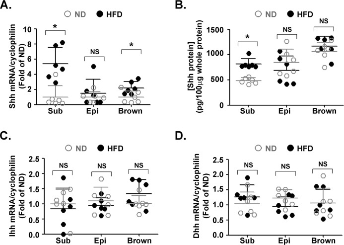
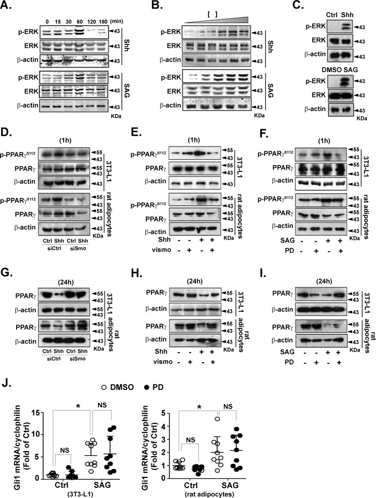
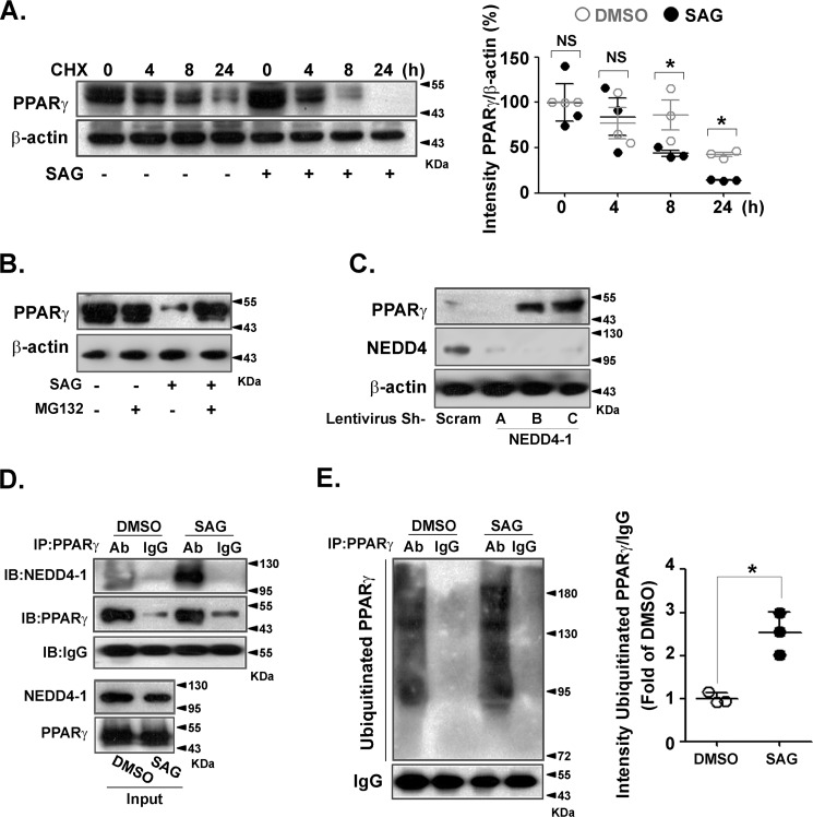
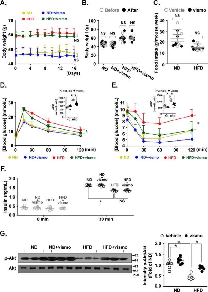
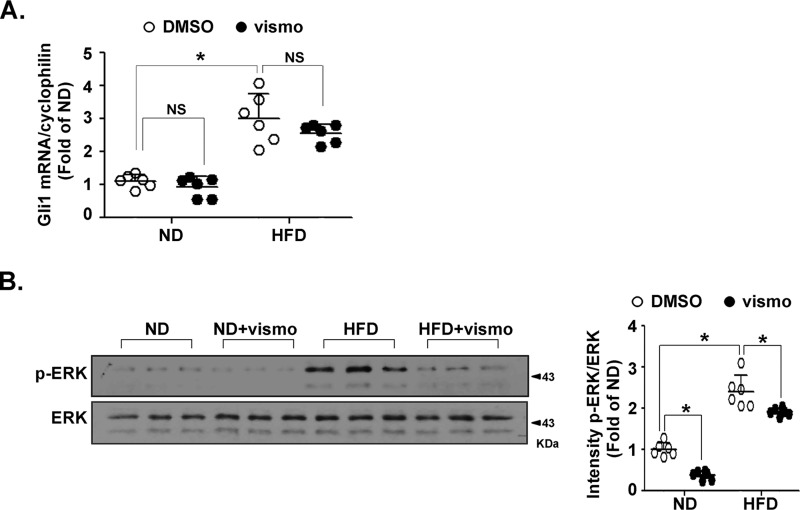
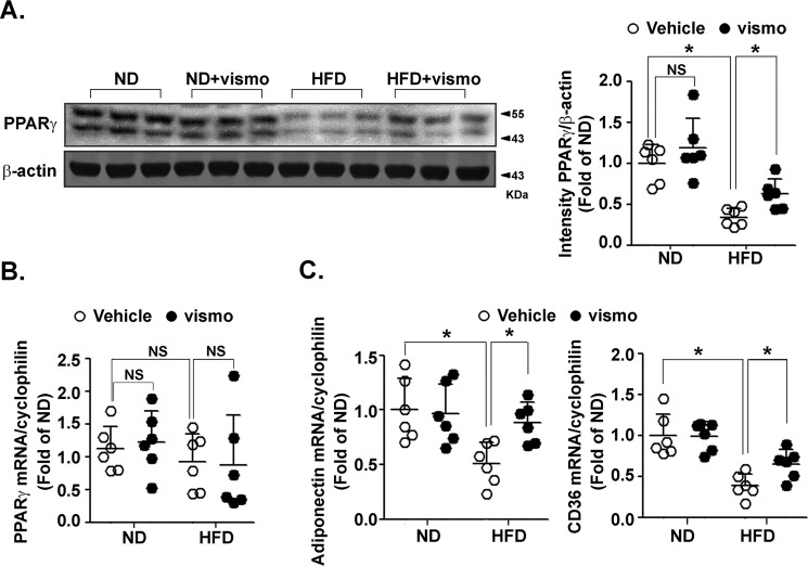
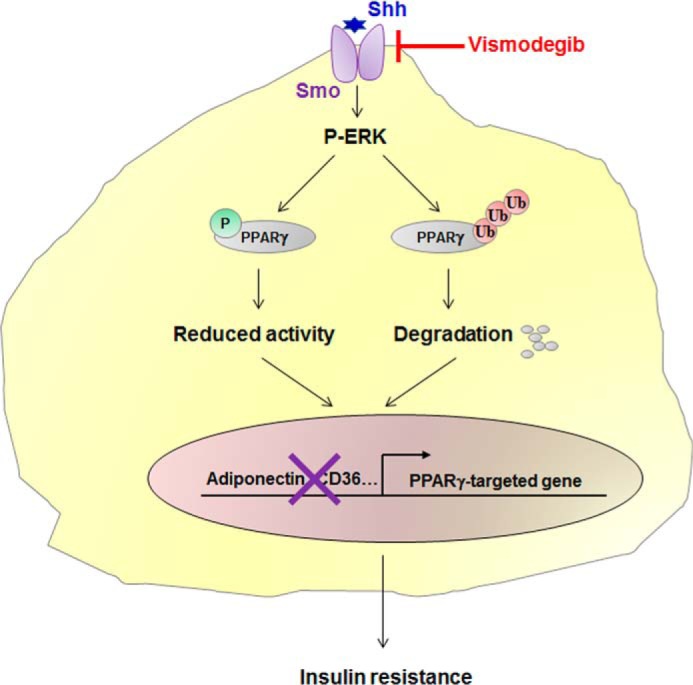
Obesity is a major risk for patients with chronic metabolic disorders including type 2 diabetes. Sonic hedgehog (Shh) is a morphogen that regulates the pancreas and adipose tissue formation during embryonic development. Peroxisome proliferator-activated receptor γ (PPARγ) is a member of the nuclear receptor superfamily and one of the most important regulators of insulin action. Here, we evaluated the role and mechanism of Shh signaling in obesity-associated insulin resistance and characterized its effect on PPARγ. We showed that Shh expression was up-regulated in subcutaneous fat from obese mice. In differentiated 3T3-L1 and primary cultured adipocytes from rats, recombinant Shh protein and SAG (an agonist of Shh signaling) activated an extracellular signal-regulated kinase (ERK)-dependent noncanonical pathway and induced PPARγ phosphorylation at serine 112, which decreased PPARγ activity. Meanwhile, Shh signaling degraded PPARγ protein via binding of PPARγ to neural precursor cell-expressed developmentally down-regulated protein 4-1 (NEDD4-1). Furthermore, vismodegib, an inhibitor of Shh signaling, attenuated ERK phosphorylation induced by a high fat diet (HFD) and restored PPARγ protein level, thus ameliorating glucose intolerance and insulin resistance in obese mice. Our finding suggests that Shh in subcutaneous fat decreases PPARγ activity and stability via activation of an ERK-dependent noncanonical pathway, resulting in impaired insulin action. Inhibition of Shh may serve as a potential therapeutic approach to treat obesity-related diabetes.

Keywords: NEDD4-1; insulin resistance; obesity; peroxisome proliferator-activated receptor (PPAR); protein degradation; sonic hedgehog (SHH); type 2 diabetes; ubiquitylation (ubiquitination).


Conflict of interest statement

The authors declare that they have no conflicts of interest with the contents of this article

Figures

Figure 1.
Figure 1.
Expression of Hh ligands in adipose tissues of obese mice. Data were obtained from mice fed with a ND or HFD for 12 weeks (n = 6 in each group). Levels of Shh mRNA (A) and protein (B) in subcutaneous (Sub), epididymal (Epi), and brown adipose tissues from lean and obese mice were measured using RT-qPCR and ELISA, respectively. Ihh (C) and Dhh (D) mRNA levels in subcutaneous, epididymal, and brown adipose tissues are shown. The results were normalized to the level of cyclophilin mRNA. *, p < 0.05; NS, no significance.
Figure 2.
Figure 2.
PPARγ protein level is decreased via the ERK pathway in adipocytes. A-C, differentiated 3T3-L1 adipocytes were serum-starved and then incubated with Shh (0.5 μg/ml) or SAG (0.5 μmol/liter) for the indicated times (A). Serum-starved 3T3-L1 adipocytes were treated with different concentrations of Shh (0, 0.1, 0.2, 0.5, 1, and 2 μg/ml) or SAG (0, 0.1, 0.2, 0.5, 1, and 2 μmol/liter) for 60 min (B). C, primary rat adipocytes (2 × 10⁵ cells/well) after serum starvation were treated with Shh (0.5 μg/ml) or SAG (0.5 μmol/liter) for 60 min. The level of p-ERK and ERK was analyzed using immunoblotting. D–F, before incubation with Shh (0.5 μg/ml) for 60 min, 3T3-L1 or primary rat adipocytes were transfected with siRNA against Smo (D) or pretreated with vismodegib (100 nmol/liter, E) for 30 min. F, PD98059 (PD, 10 μmol/liter)-pretreated cells were exposed to SAG (0.5 μmol/liter) for 60 min. Levels of p-PPARγ and PPARγ was analyzed by using immunoblotting. G–I, Smo siRNA-transfected (G) or vismodegib-pretreated (H) cells were incubated with Shh (0.5 μg/ml) for 24 h. I, PD98059 (PD, 10 μmol/liter)-pretreated cells were exposed to SAG (0.5 μmol/liter) for 24 h. Levels of PPARγ and β-actin were analyzed using immunoblotting. Immunoblots shown are representative of 3 independent experiments. J, differentiated 3T3-L1 (left) and primary rat adipocytes (right) were pretreated with or without PD98059 (PD, 10 μmol/liter) for 30 min, and then exposed to SAG (0.5 μmol/liter) for 24 h. Cell lysates were analyzed to determine the mRNA level of Gli1. The results were normalized to the level of cyclophilin mRNA. Data were from 3 independent experiments performed in triplicate. *, p < 0.05; NS, no significance.
Figure 3.
Figure 3.
Shh signaling decreased PPARγ stability via NEDD4-1–dependent ubiquitination. HEK 293 cells were transfected with PPARγ overexpression plasmid. A, cells were pretreated with cyclohexamide (CHX) (5 μg/ml) for 30 min before exposure to SAG (0.5 μmol/liter), followed by immunoblotting to detect PPARγ and β-actin (left). Quantifications of band intensity normalized to β-actin (right). B, cells were incubated with SAG (0.5 μmol/liter) for 24 h in the presence or absence of MG132 (10 μmol/liter) pretreatment. C, cells were infected with scramble shRNA lentivirus or different lentiviral shRNA constructs against NEDD4-1. PPARγ, NEDD4-1, and β-actin levels were analyzed by immunoblotting. D, immunoblotting (IB) of whole cell lysates (Input) and immunoprecipitates (IP) from PPARγ-overexpressing HEK 293 cells with or without SAG (0.5 μmol/liter) treatment for 24 h. MG132 (10 μmol/liter) was added to the medium 12 h before collecting. E, PPARγ-transfected HEK 293 cells were pretreated with MG132 (10 μmol/liter) and then incubated with or without SAG (0.5 μmol/liter) for 24 h. PPARγ was immunoprecipitated from cell lysates and immunoblotted with an anti-ubiquitin antibody (left). Band intensities were normalized to that of IgG (right). Immunoblots shown are representative of 3 independent experiments. *, p < 0.05.
Figure 4.
Figure 4.
Vismodegib improved insulin resistance in obese mice. Mice fed a ND or HFD for 12 weeks (n = 6 in each group) were injected with vismodegib every other day at a dose of 5 mg/kg. A, body weight was measured before every injection. NS: ND V.S. ND+vismo, NS: HFD V.S. HFD+vismo. B, body weights of ND- or HFD-fed mice before and after vismodegib treatment. C, level of average food intake during vismodegib treatment. Glucose tolerance test (D) and insulin tolerance test (E) of ND- or HFD-fed mice with or without vismodegib treatment. *, HFD V.S. HFD+vismo. Areas under the curve (AUC) were determined. F, level of serum insulin in ND- or HFD-fed mice with vismodegib treatment before and after insulin injection (30 min). G, protein levels of p-Akt and Akt in subcutaneous adipose tissue were measured by immunoblotting. Quantifications of band intensity was normalized to β-actin (right). F, tail vein blood was sampled before and 30 min after glucose injection for the measurement of insulin level. *, p < 0.05; NS, no significance.
Figure 5.
Figure 5.
Vismodegib treatment affected the ERK pathway in obese mice. Mice fed a ND or HFD for 12 weeks (n = 6 in each group) were injected with vismodegib every other day at a dose of 5 mg/kg. After sacrifice, the subcutaneous adipose tissues were immediately dissected and underwent quick-freezing in liquid nitrogen. A, level of Gli1 mRNA was measured by RT-qPCR. The result was normalized to the level of cyclophilin. B, protein levels of p-ERK and ERK in subcutaneous adipose tissue were measured by immunoblotting. Quantification of band intensity normalized to β-actin (right). *, p < 0.05; NS, no significance.
Figure 6.
Figure 6.
Vismodegib restored PPARγ protein levels in subcutaneous fat of obese mice. Mice fed a ND or HFD for 12 weeks (n = 6 in each group) were injected with vismodegib every other day at a dose of 5 mg/kg. After sacrifice, the subcutaneous adipose tissues were immediately dissected and underwent quick-freezing in liquid nitrogen. The protein level of PPARγ in subcutaneous adipose tissue was measured by immunoblotting (left). Quantification of band intensity normalized to β-actin (right). The mRNA levels of PPARγ (B) and PPARγ-targeted genes (C) were measured by RT-qPCR. The results were normalized to the level of cyclophilin. *, p < 0.05; NS, no significance.
Figure 7.
Figure 7.
Role of Shh signaling in obesity-induced insulin resistance. Increased Shh expression in subcutaneous fat of obese mice activates p-ERK, which phosphorylates PPARγ at serine 112, leading to an impaired PPARγ activity. On the other hand, Shh-activated ERK phosphorylation promotes NEDD4-1–dependent PPARγ ubiquitination and degradation. Transcription of PPARγ-targeted genes is therefore reduced, leading to an impaired insulin action. Thus, by targeting the Shh receptor Smo, vismodegib instigates HFD–induced insulin resistance in obese mice.

Similar articles

Cited by

References

    1. Tokarz V. L., MacDonald P. E., and Klip A. (2018) The cell biology of systemic insulin function. J. Cell Biol. 217, 2273–2289 10.1083/jcb.201802095 - DOI - PMC - PubMed
    1. Page M. M., and Johnson J. D. (2018) Mild suppression of hyperinsulinemia to treat obesity and insulin resistance. Trends Endocrinol. Metab. 29, 389–399 10.1016/j.tem.2018.03.018 - DOI - PubMed
    1. Koren D., and Taveras E. M. (2018) Association of sleep disturbances with obesity, insulin resistance and the metabolic syndrome. Metabolism 84, 67–75 10.1016/j.metabol.2018.04.001 - DOI - PubMed
    1. Tchernof A., and Després J. P. (2013) Pathophysiology of human visceral obesity: an update. Physiol. Rev. 93, 359–404 10.1152/physrev.00033.2011 - DOI - PubMed
    1. Abate N., Garg A., Peshock R. M., Stray-Gundersen J., and Grundy S. M. (1995) Relationships of generalized and regional adiposity to insulin sensitivity in men. J. Clin. Investig. 96, 88–98 10.1172/JCI118083 - DOI - PMC - PubMed

Publication types

MeSH terms

LinkOut - more resources