. 2019 Feb 15;294(7):2267-2278.
doi: 10.1074/jbc.RA118.004383. Epub 2018 Dec 20.

Estrogen receptor α promotes protein synthesis by fine-tuning the expression of the eukaryotic translation initiation factor 3 subunit f (eIF3f)

Affiliations

Estrogen receptor α promotes protein synthesis by fine-tuning the expression of the eukaryotic translation initiation factor 3 subunit f (eIF3f)

Rafael Cuesta et al. J Biol Chem. .

Abstract

Approximately two thirds of all breast cancer cases are estrogen receptor (ER)-positive. The treatment of this breast cancer subtype with endocrine therapies is effective in the adjuvant and recurrent settings. However, their effectiveness is compromised by the emergence of intrinsic or acquired resistance. Thus, identification of new molecular targets can significantly contribute to the development of novel therapeutic strategies. In recent years, many studies have implicated aberrant levels of translation initiation factors in cancer etiology and provided evidence that identifies these factors as promising therapeutic targets. Accordingly, we observed reduced levels of the eIF3 subunit eIF3f in ER-positive breast cancer cells compared with ER-negative cells, and determined that low eIF3f levels are required for proper proliferation and survival of ER-positive MCF7 cells. The expression of eIF3f is tightly controlled by ERα at the transcriptional (genomic pathway) and translational (nongenomic pathway) level. Specifically, estrogen-bound ERα represses transcription of the EIF3F gene, while promoting eIF3f mRNA translation. To regulate translation, estrogen activates the mTORC1 pathway, which enhances the binding of eIF3 to the eIF4F complex and, consequently, the assembly of the 48S preinitiation complexes and protein synthesis. We observed preferential translation of mRNAs with highly structured 5'-UTRs that usually encode factors involved in cell proliferation and survival (e.g. cyclin D1 and survivin). Our results underscore the importance of estrogen-ERα-mediated control of eIF3f expression for the proliferation and survival of ER-positive breast cancer cells. These findings may provide rationale for the development of new therapies to treat ER-positive breast cancer.

Keywords: ERα; breast cancer; eIF3f; estrogen receptor; eukaryotic translation initiation; gene expression; mTORC1; translation.


Conflict of interest statement

The authors declare that they have no conflicts of interest with the contents of this article

Figures

Figure 1.
Figure 1.
Expression of eIF3a, eIF3b, and eIF3f subunits in a panel of breast cancer cells. A, ER-positive (MCF7, T47D, ZR75.1, and MDA-MB-361) and ER-negative (BT-474, MDA-MB-231, and MDA-MB-436) cells (2.5 × 10⁵ cells per well in 6-well plates) were grown in complete media until 80% confluency, whole-cell extracts were obtained, and indicated proteins were analyzed by immunoblotting. Representative blots are shown. B, intensities of eIF3a, eIF3b, eIF3f, and actin bands from four biological replicates were quantified with Image Studio Software 5.2. The values for the eIF3 subunits were normalized to actin values and plotted as mean ± S.E. (*, p ≤ 0.05; **, p ≤ 0.01).
Figure 2.
Figure 2.
Estrogen reduces transcription of the EIF3F gene. A, ER-positive MCF7 cells were grown in phenol red–free DMEM supplemented with 10% charcoal-stripped FBS (low-estrogen medium) for 3 days before stimulation with vehicle (EtOH) or estradiol (10 nm) for the indicated times. Total RNA was purified and the levels of TFF1, eIF3f, and GAPDH mRNAs were determined by RT-qPCR. TFF1 and eIF3f values were normalized to GAPDH and mean ± S.E. of three independent experiments were expressed relative to vehicle-treated sample at time 0 (set to 1) (*, p ≤ 0.05; **, p ≤ 0.01). B, MCF7 cells were grown in low-estrogen medium for 3 days and then stimulated with vehicle (EtOH) or estradiol (10 nm) for 12 h before adding actinomycin D (5 μg/ml) and continuing the incubation for indicated times. TFF1 and eIF3f mRNAs were analyzed as described in A. Data represented as mean ± S.E. of three independent experiments (*, p ≤ 0.05). C, MCF7 and MDA-MB-231 cells were grown in low-estrogen medium for 3 days before stimulation with vehicle (EtOH), estradiol (10 nm), or tamoxifen (100 nm) for 24 h. TFF1 and eIF3f mRNAs were purified and analyzed as described in A. Mean ± S.E. of three independent experiments were plotted (*, p ≤ 0.05; **, p ≤ 0.01).
Figure 3.
Figure 3.
ERα mediates transcriptional down-regulation of eIF3f expression in estrogen-stimulated cells. A, MCF7 cells transfected with scrambled siRNA or siRNA against ERα were grown in low-estrogen medium for 3 days before stimulation with vehicle (EtOH) or estradiol (10 nm) for 24 h. Total RNA was purified and the levels of TFF1, eIF3f, and GAPDH mRNAs were determined by RT-qPCR. TFF1 and eIF3f values were normalized by GAPDH and expressed relative to scrambled siRNA-transfected cells treated with vehicle (set to 1). Data represented as mean ± S.E. of five independent experiments (*, p ≤ 0.05; **, p ≤ 0.01). B, MCF7 cells were transfected and treated as in A. Whole-cell extracts were prepared and indicated proteins were analyzed by immunoblotting.
Figure 4.
Figure 4.
ERα may repress EIF3F transcription by a mechanism of physiologic squelching. A, MCF7 cells grown in low-estrogen medium for 3 days were treated with cycloheximide (10 μg/ml) for 1 h before stimulation with vehicle (EtOH) or estradiol (10 nm) for 24 h. The levels of TFF1 and eIF3f mRNAs were determined as described in Fig. 3A. Data from four independent experiments were represented as mean ± S.E. (*, p ≤ 0.05; **, p ≤ 0.01; ***, p ≤ 0.001). B, MCF7 cells were estrogen-deprived for 3 days and then treated with vehicle (EtOH) or estradiol (10 nm) for 30 min. Nuclear extracts were obtained and the binding of ERα or RNA Pol II to EIF3F promoter or distal estrogen response element (ERE) was determined by ChIP assays using indicated set of primers. Values indicate percentage of input DNA bound to ERα or Pol II after normalizing to control IgG values. Mean ± S.E. of four independent experiments were graphed. C, binding of ERα or RNA Pol II to TFF1 promoter was determined as in D (*, p ≤ 0.05).
Figure 5.
Figure 5.
Estrogen-ERα pathway facilitates the binding of eIF3 to eIF4F and promotes cap-dependent translation. A, MCF7 cells transfected with R-HCV-L bicistronic plasmid were grown in phenol red–free DMEM containing 5% charcoal-treated FBS for 3 days before being treated with vehicle, estradiol (100 nm), estradiol and rapamycin (20 nm), estradiol and fulvestrant (100 nm), or tamoxifen (100 nm) for 24 h. Cell extracts were obtained, and Renilla and Firefly Luciferase activities were determined. Renilla/Firefly ratios were calculated and mean ± S.E. of three independent experiments were plotted relative to vehicle-treated cells (set to 1) (*, p ≤ 0.05). B, MCF7 cells were estrogen-deprived for 3 days. Cells were treated with vehicle, rapamycin (20 nm), or fulvestrant (100 nm) for 30 min before being stimulated with estradiol (100 nm) or tamoxifen (100 nm) for 2 h. Cell extracts were obtained and translation initiation complexes isolated using m7GTP-agarose beads. Indicated proteins were analyzed by immunoblotting. C, cell extracts obtained as in B were incubated with anti-eIF3b antibody overnight at 4 °C. Purified immunocomplexes were resolved by SDS-PAGE and indicated proteins were analyzed by immunoblotting. D, MCF7 cells were cultured and treated for 24 h as in B. Cell extracts were prepared and resolved by SDS-PAGE, and indicated proteins were detected by immunoblotting. E, MCF7 cells were grown in phenol red–free media supplemented with 5% charcoal-stripped FBS for 3 days before stimulation with vehicle (EtOH), estradiol (100 nm), estradiol and mTOR inhibitor pp242 (2.5 μm), or tamoxifen (100 nm) for 24 or 36 h. Cell lysates were prepared and indicated proteins were analyzed by immunoblotting.
Figure 6.
Figure 6.
Estrogen induces the dissociation of ERα from eIF3 and the nuclear localization of ERα. A, HA-transfected or HA-eIF3f-transfected MCF7 cells were incubated in low-estrogen DMEM for 3 days, followed by stimulation with estrogen (10 nm) for 30 min. Cell extracts were prepared, precleared with protein G–agarose beads for 1 h at 4 °C, and incubated with anti-HA antibody overnight at 4 °C. Isolated immunocomplexes were resolved by SDS-PAGE and indicated proteins were detected by immunoblotting. B and C, MCF7 cells were treated as in A, and cell extracts were precleared with protein A/G–agarose for 1 h at 4 °C followed by incubation with anti-ERα (B) or anti-eIF3b (C) antibodies overnight at 4 °C. Isolated immunocomplexes were resolved by SDS-PAGE and indicated proteins were analyzed by immunoblotting. D, MCF7 cells were estrogen-deprived for 3 days, followed by stimulation with estrogen (10 nm) for 1 h. Nuclear and cytoplasmic extracts were generated as described in “Experimental procedures” and separated by SDS-PAGE. Indicated proteins were analyzed by immunoblotting.
Figure 7.
Figure 7.
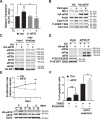
eIF3F overexpression prevents estrogen-induced up-regulation of cap-dependent translation. A, MCF7 cells transfected with empty vector or an eIF3f-expressing plasmid and R-HCV-L bicistronic plasmid were grown in phenol red–free DMEM containing 5% charcoal-treated FBS for 3 days before being treated with vehicle or estradiol (100 nm) for 24 h. Cells extracts were obtained and Renilla and Firefly Luciferase activities were determined. Renilla/Firefly ratios were calculated and values plotted relative to untreated control cells (*, p ≤ 0.05). B, MCF7 cells were transfected with plasmids expressing HA tag or HA-eIF3f and grown in phenol red–free DMEM containing 5% charcoal-treated FBS for 2 days. Then, cells were treated with vehicle (EtOH) or estrogen (100 nm) for 24 h. Whole-cell extracts were obtained and resolved by SDS-PAGE, and indicated proteins analyzed by immunoblotting. C, MCF7 cells were cotransfected with pcDNA3 and empty vector or an eIF3f-expressing plasmid (pcDNA3 provides neomycin resistance to transfected cells). After 24 h, cells were selected in low-estrogen medium containing G410 (0.6 mg/ml) for 48 h and then stimulated with estrogen (100 nm) for 2 h. Whole-cell extracts were prepared and cap-binding complexes were isolated and resolved by SDS-PAGE; indicated proteins were analyzed by immunoblotting. D, cell extracts prepared as in C were used to pull down eIF3b protein. Immunoprecipitated proteins were separated by SDS-PAGE and indicated proteins analyzed by immunoblotting. (Band corresponding to heavy IgG chain is marked with an asterisk). E, MCF7 cells were transfected as in B. After 24 h, cells were seeded into 96-well plates and incubated in complete medium for additional 24 h. Then, transfected cells were grown in phenol red–free DMEM containing 5% charcoal-treated FBS and estrogen (100 nm) for indicated days. Viable cells were estimated using MTT cell viability assays, and values represented as mean ± S.E. relative to day 0 cells (set to 1) determined from three independent assays (*, p ≤ 0.05; **, p ≤ 0.01). The blots show the levels of the endogenous and ectopic eIF3f at the different time points (C, Ctrl; 3f, HA-eIF3f). F, MCF7 cells were transfected as in C and selected in DMEM containing 10% FBS and G418 for 48 h, as indicated. Cells were treated with vehicle (DMSO) or etoposide (50 μm) for 24 h, as indicated, and apoptotic cells were labeled with Guava Nexin Reagent and quantified using the Guava easyCyte Flow Cytometer. Percentage of apoptotic cells was represented as mean ± S.E. from three independent experiments (*, p ≤ 0.05).

Similar articles

Cited by

References

    1. Miller T. W., Balko J. M., and Arteaga C. L. (2011) Phosphatidylinositol 3-kinase and antiestrogen resistance in breast cancer. J. Clin. Oncol. 29, 4452–4461 10.1200/JCO.2010.34.4879 - DOI - PMC - PubMed
    1. De Marchi T., Foekens J. A., Umar A., and Martens J. W. (2016) Endocrine therapy resistance in estrogen receptor (ER)-positive breast cancer. Drug Discov. Today 21, 1181–1188 10.1016/j.drudis.2016.05.012 - DOI - PubMed
    1. Miller W. R., and Larionov A. A. (2012) Understanding the mechanisms of aromatase inhibitor resistance. Breast Cancer Res. 14, 201 10.1186/bcr2931 - DOI - PMC - PubMed
    1. Rondón-Lagos M., Villegas V. E., Rangel N., Sánchez M. C., and Zaphiropoulos P. G. (2016) Tamoxifen resistance: Emerging molecular targets. Int. J. Mol. Sci. 17, 1357 10.3390/ijms17081357 - DOI - PMC - PubMed
    1. Carroll J. S., Meyer C. A., Song J., Li W., Geistlinger T. R., Eeckhoute J., Brodsky A. S., Keeton E. K., Fertuck K. C., Hall G. F., Wang Q., Bekiranov S., Sementchenko V., Fox E. A., Silver P. A., Gingeras T. R., Liu X. S., and Brown M. (2006) Genome-wide analysis of estrogen receptor binding sites. Nat. Genet. 38, 1289–1297 10.1038/ng1901 - DOI - PubMed

Publication types

MeSH terms