Review
. 2019 Apr;20(4):221-234.
doi: 10.1038/s41576-018-0087-x.

Mechanisms regulating zygotic genome activation

Affiliations
Review

Mechanisms regulating zygotic genome activation

Katharine N Schulz et al. Nat Rev Genet. 2019 Apr.

Abstract

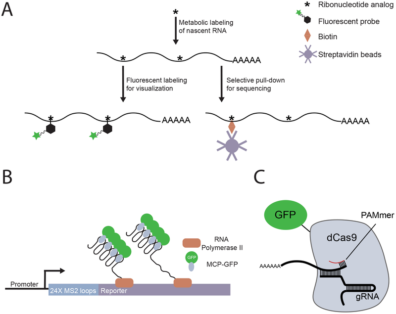
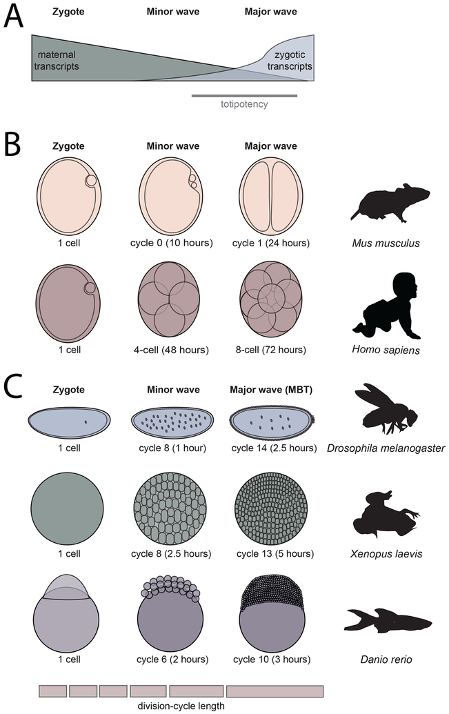
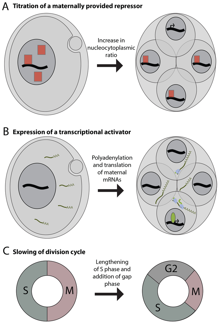
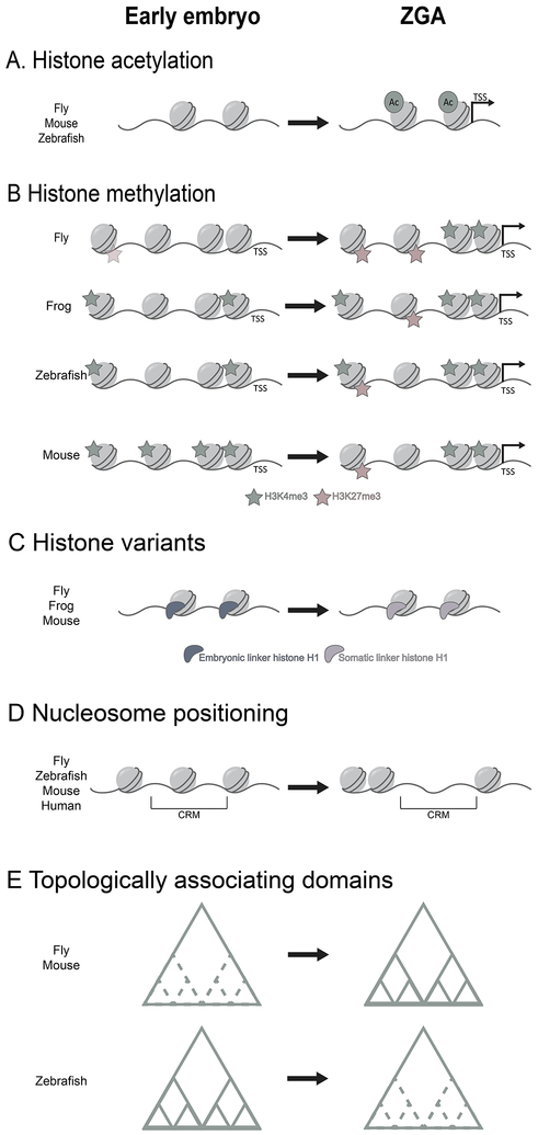
Following fertilization, the two specified gametes must unite to create an entirely new organism. The genome is initially transcriptionally quiescent, allowing the zygote to be reprogrammed into a totipotent state. Gradually, the genome is activated through a process known as the maternal-to-zygotic transition, which enables zygotic gene products to replace the maternal supply that initiated development. This essential transition has been broadly characterized through decades of research in several model organisms. However, we still lack a full mechanistic understanding of how genome activation is executed and how this activation relates to the reprogramming of the zygotic chromatin architecture. Recent work highlights the central role of transcriptional activators and suggests that these factors may coordinate transcriptional activation with other developmental changes.


Figures

Figure 1:
Figure 1:. New technologies enable precise detection of zygotic genome activation
(A)Detection of nascent transcripts by metabolic labeling: Cells are supplied with ribonucleoside or ribonucleotide analogs (for example, 5-ethynyl uridine or 4-thio-UTP) to label actively transcribed RNA. The labeled RNA is functionalized through coupling to a biophysical probe, such as a fluorescent azide for visualization or a biotin group for selective pull-down and sequencing. (B) Detection of nascent transcripts using MS2-based reporters: A series of MS2 sequences is introduced adjacent to a gene of interest. As the MS2 motifs are transcribed they form RNA stem loops that are bound by a maternally provided MS2 coat protein fused to fluorescent protein (MCP-GFP) to collectively produce a fluorescent spot. (C) Detection of transcripts by RNA-targeted deadCas9 (dCas9) fused to a fluorescent protein (for example, GFP): The dCas9-GFP fusion protein is targeted to RNA through interaction with a guide RNA (gRNA), which contains sequence that is complementary to the RNA of interest, and a DNA oligo that contains the protospacer adjacent motif (PAM), known as a PAMmer.
Figure 2.
Figure 2.. Zygotic genome activation is conserved across animals.
(A) In the first hours of life, animals undergo a process called the maternal-to-zygotic transition (MZT) in which the clearance of maternal products is coordinated with the activation of zygotic transcription. A totipotent state (gray bar) is established during this transition. (B,C) Key stages of zygotic genome activation are outlined for five model species, indicated on the right. The absolute time (in hours post fertilization) is indicated below. All species begin life as a single-cell zygote. Zygotic transcription initiates in an early minor wave, which is later followed by a major wave of genome activation. (B) In mice and humans, early cell divisions do not occur as rapidly as in externally fertilized organisms such as frogs, zebrafish and flies (C). Nonetheless, as in other species, genome activation is a gradual process with a minor wave and major wave of transcription. (C) In frogs, zebrafish, and flies, the rapid division cycles that characterize early development gradually slow over the course of the MZT. In these species, the major wave of genome activation coincides with the mid-blastula transition (MBT). The MBT involves the end of synchronous division cycles, the introduction of a gap phase (G2) to the cell cycle, and additional, species-specific developmental changes.
Figure 3.
Figure 3.. Several mechanisms contribute to the timing of zygotic genome activation.
(A) A maternally supplied repressor (red square) prevents transcription in the early embryo. As the ratios of genetic material (black line) or nuclear volume (grey circle) to cytoplasm increase with each cell division, the repressor is titrated and transcription initiates in cells in which repressor concentration has fallen below a threshold level (B) The early embryo lacks a key transcriptional activator (green oval). Polyadenylation and translation of maternally supplied mRNA leads to its accumulation. Once a threshold level has been reached, the factor enables expression of its target genes. (C) The rapid early cell cycles consist of only a DNA replication (S) phase and mitosis (M). At the major wave of zygotic genome activation, the cell cycle slows and a gap phase (G2) is introduced, reducing the time restraint initially placed on transcription.
Figure 4.
Figure 4.. Chromatin is reprogrammed during zygotic genome activation.
(A) In flies, mice, and zebrafish, histone acetylation increases over the course of ZGA, marking genes for activation during this transition. TSS, transcription start site. (B) In flies, the early embryo may contain low levels of H3K27me3, and both H3K27me3 and H3K4me3 increase sharply during the major wave of genome activation. In frogs, H3K4me3 is present in the early embryo at low levels and increases over the course of ZGA. H3K27me3 is established later, during the major wave of transcription. In early zebrafish, H3K4me3 appears to poise genes for activation. During the major wave of ZGA, H3K4me3 levels increase and H3K27me3 is established. H3K27me3 co-marks histones with H3K4me3, forming bivalent domains. In mice, unusual broad domains of H3K4me3 are restricted to TSS-associated peaks during ZGA with H3K27me3 established later. (C) In flies, frogs, and mice, embryonic variants of linker histone H1 are replaced with their somatic counterparts at the major onset of ZGA. In flies, it has been shown that incorporation of the somatic H1 variant is instrumental for genome activation to occur. (D) Defined cis-regulatory elements (CRM), characterized by open chromatin, are established during ZGA in flies, zebrafish, mice, and humans. (E) In flies and mice, the boundaries of topologically associating domains (TADs) are established concurrently with ZGA. In zebrafish, TADs are present in the early embryo but are lost prior to the major wave of genome activation.

References

    1. Gurdon JB The Developmental Capacity of Nuclei taken from Intestinal Epithelium Cells of Feeding Tadpoles. Development 10, (1962). - PubMed
    1. Campbell KHS, McWhir J, Ritchie WA & Wilmut I Sheep cloned by nuclear transfer from a cultured cell line. Nature 380, 64–66 (1996). - PubMed
    1. Newport J & Kirschner M A major developmental transition in early Xenopus embryos: II. Control of the onset of transcription. Cell 30, 687–696 (1982). - PubMed
    1. Tadros W & Lipshitz HD The maternal-to-zygotic transition: a play in two acts. Development 136, 3033–3042 (2009). - PubMed
    1. Yartseva V & Giraldez AJ The Maternal-to-Zygotic Transition During Vertebrate Development: A Model for Reprogramming. Curr. Top. Dev. Biol. 113, 191–232 (2015). - PMC - PubMed

Publication types

MeSH terms