

Spectrum of tau pathologies in Huntington's disease
- PMID: 30573872
- PMCID: PMC9342691
- DOI: 10.1038/s41374-018-0166-9
Spectrum of tau pathologies in Huntington's disease
Abstract
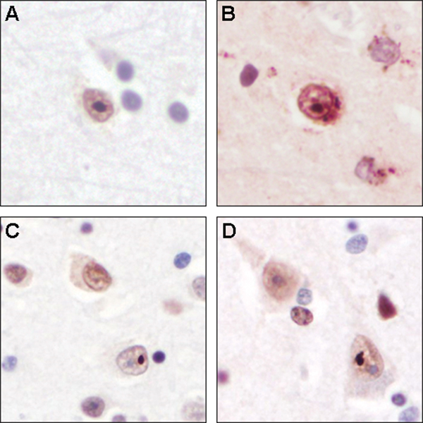
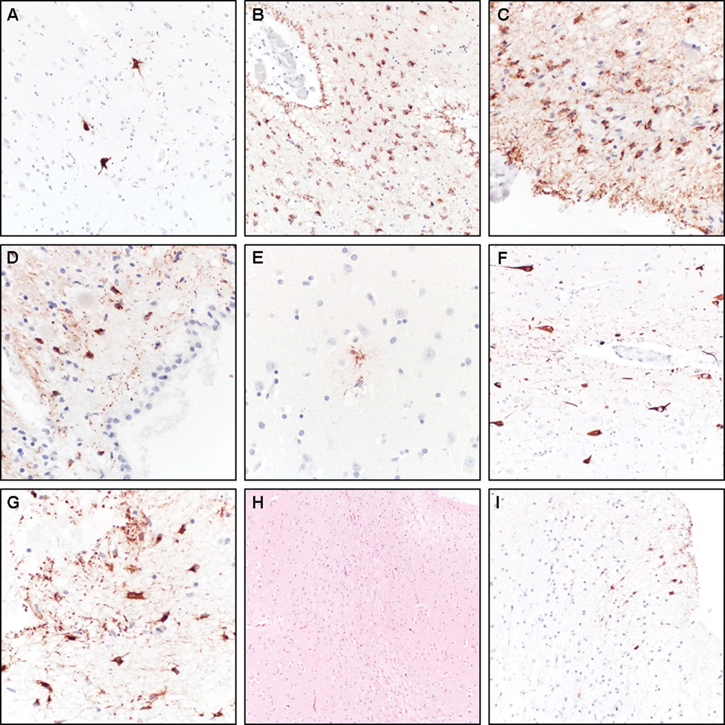
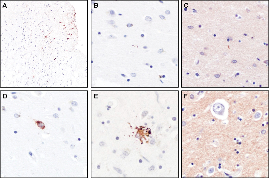
Huntington's disease (HD) is an autosomal dominant disorder caused by a trinucleotide expansion in the huntingtin gene. Recently, a new role for tau has been implicated in the pathogenesis of HD, whereas others have argued that postmortem tau pathology findings are attributable to concurrent Alzheimer's disease pathology. The frequency of other well-defined common age-related tau pathologies in HD has not been examined in detail. In this single center, retrospective analysis, we screened seven cases of Huntington's disease (5 females, 2 males, age at death: 47-73 years) for neuronal and glial tau pathology using phospho-tau immunohistochemistry. All seven cases showed presence of neuronal tau pathology. Five cases met diagnostic criteria for primary age-related tauopathy (PART), with three cases classified as definite PART and two cases as possible PART, all with a Braak stage of I. One case was diagnosed with low level of Alzheimer's disease neuropathologic change. In the youngest case, rare perivascular aggregates of tau-positive neurons, astrocytes and processes were identified at sulcal depths, meeting current neuropathological criteria for stage 1 chronic traumatic encephalopathy (CTE). Although the patient had no history of playing contact sports, he experienced several falls, but no definitive concussions during his disease course. Three of the PART cases and the CTE-like case showed additional evidence of aging-related tau astrogliopathy. None of the cases showed significant tau pathology in the striatum. In conclusion, while we found evidence for tau hyperphosphorylation and aggregation in all seven of our HD cases, the tau pathology was readily classifiable into known diagnostic entities and most likely represents non-specific age- or perhaps trauma-related changes. As the tau pathology was very mild in all cases and not unexpected for a population of this age range, it does not appear that the underlying HD may have promoted or accelerated tau accumulation.
Conflict of interest statement
Disclosure/Conflict of Interest
None of the authors has conflicts of interest to disclose.
Figures
Similar articles
-
Tau immunophenotypes in chronic traumatic encephalopathy recapitulate those of ageing and Alzheimer's disease.Brain. 2020 May 1;143(5):1572-1587. doi: 10.1093/brain/awaa071. Brain. 2020. PMID: 32390044 Free PMC article.
-
Is Huntington's disease a tauopathy?Brain. 2016 Apr;139(Pt 4):1014-25. doi: 10.1093/brain/aww021. Epub 2016 Mar 11. Brain. 2016. PMID: 26969684 Review.
-
Tau hyperphosphorylation and deregulation of calcineurin in mouse models of Huntington's disease.Hum Mol Genet. 2015 Jan 1;24(1):86-99. doi: 10.1093/hmg/ddu456. Epub 2014 Sep 8. Hum Mol Genet. 2015. PMID: 25205109
-
Implications of Tau Dysregulation in Huntington's Disease and Potential for New Therapeutics.J Huntingtons Dis. 2023;12(1):1-13. doi: 10.3233/JHD-230569. J Huntingtons Dis. 2023. PMID: 37092231 Free PMC article. Review.
-
Detection of astrocytic tau pathology facilitates recognition of chronic traumatic encephalopathy neuropathologic change.Acta Neuropathol Commun. 2022 Apr 11;10(1):50. doi: 10.1186/s40478-022-01353-4. Acta Neuropathol Commun. 2022. PMID: 35410438 Free PMC article.
Cited by
-
Huntington disease: Advances in the understanding of its mechanisms.Clin Park Relat Disord. 2020 May 6;3:100056. doi: 10.1016/j.prdoa.2020.100056. eCollection 2020. Clin Park Relat Disord. 2020. PMID: 34316639 Free PMC article. Review.
-
Chronic Traumatic Encephalopathy and Neuropathological Comorbidities.Semin Neurol. 2020 Aug;40(4):384-393. doi: 10.1055/s-0040-1713628. Epub 2020 Jun 30. Semin Neurol. 2020. PMID: 32629520 Free PMC article. Review.
-
Neuropathology and pathogenesis of extrapyramidal movement disorders: a critical update. II. Hyperkinetic disorders.J Neural Transm (Vienna). 2019 Aug;126(8):997-1027. doi: 10.1007/s00702-019-02030-y. Epub 2019 Jun 24. J Neural Transm (Vienna). 2019. PMID: 31236685 Review.
-
Age-Dependent Increase in Tau Phosphorylation at Serine 396 in Huntington's Disease Prefrontal Cortex.J Huntingtons Dis. 2023;12(3):267-281. doi: 10.3233/JHD-230588. J Huntingtons Dis. 2023. PMID: 37694372 Free PMC article.
-
Huntington disease: new insights into molecular pathogenesis and therapeutic opportunities.Nat Rev Neurol. 2020 Oct;16(10):529-546. doi: 10.1038/s41582-020-0389-4. Epub 2020 Aug 14. Nat Rev Neurol. 2020. PMID: 32796930 Review.
References
-
- A novel gene containing a trinucleotide repeat that is expanded and unstable on Huntington’s disease chromosomes. The Huntington’s Disease Collaborative Research Group. Cell 1993;72:971–983. - PubMed
-
- Kim SD, Fung VS. An update on Huntington’s disease: from the gene to the clinic. Current opinion in neurology 2014;27:477–483. - PubMed
-
- Sturrock A, Leavitt BR. The clinical and genetic features of Huntington disease. Journal of geriatric psychiatry and neurology 2010;23:243–259. - PubMed
-
- Vonsattel JP, DiFiglia M. Huntington disease. J Neuropathol Exp Neurol 1998;57:369–384. - PubMed
-
- Vonsattel JP, Myers RH, Stevens TJ, et al. Neuropathological classification of Huntington’s disease. J Neuropathol Exp Neurol 1985;44:559–577. - PubMed
Publication types
MeSH terms
Substances
Grants and funding
LinkOut - more resources
Full Text Sources
Medical
Research Materials
Miscellaneous
