Review
. 2018 Dec 6:12:483.
doi: 10.3389/fncel.2018.00483. eCollection 2018.

The Emerging Role of Mechanics in Synapse Formation and Plasticity

Affiliations
Review

The Emerging Role of Mechanics in Synapse Formation and Plasticity

Devrim Kilinc. Front Cell Neurosci. .

Abstract

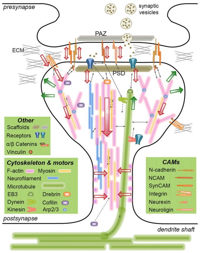
The regulation of synaptic strength forms the basis of learning and memory, and is a key factor in understanding neuropathological processes that lead to cognitive decline and dementia. While the mechanical aspects of neuronal development, particularly during axon growth and guidance, have been extensively studied, relatively little is known about the mechanical aspects of synapse formation and plasticity. It is established that a filamentous actin network with complex spatiotemporal behavior controls the dendritic spine shape and size, which is thought to be crucial for activity-dependent synapse plasticity. Accordingly, a number of actin binding proteins have been identified as regulators of synapse plasticity. On the other hand, a number of cell adhesion molecules (CAMs) are found in synapses, some of which form transsynaptic bonds to align the presynaptic active zone (PAZ) with the postsynaptic density (PSD). Considering that these CAMs are key components of cellular mechanotransduction, two critical questions emerge: (i) are synapses mechanically regulated? and (ii) does disrupting the transsynaptic force balance lead to (or exacerbate) synaptic failure? In this mini review article, I will highlight the mechanical aspects of synaptic structures-focusing mainly on cytoskeletal dynamics and CAMs-and discuss potential mechanoregulation of synapses and its relevance to neurodegenerative diseases.

Keywords: cell adhesion molecules; cytoskeleton; dendritic spine; mechanotransduction; motor proteins; synaptic scaffold proteins.


Figures

Figure 1
Figure 1
Mechanically-relevant components of an excitatory synapse. Presynaptic vesicle fusion machinery and postsynaptic receptors are held in place by their respective scaffold proteins, which are physically linked to cell adhesion molecules (CAMs) and the cytoskeleton. Direct interactions are indicated with thin, continuous arrows. Translational movements are indicated with thin, broken arrows. Only postsynaptic cytoskeleton is depicted for simplicity. Major cytoskeletal filaments (actin, neurofilaments, microtubules) and a select set of associated molecules (microtubule end-binding protein EB3, actin severing/stabilizing protein cofilin, actin branch-inducing Arp2/3 complex) are depicted. Spine base and center are occupied by stable, dense F-actin, whereas the periphery is occupied by dynamic, branched F-actin. F-actin rings line the spine shaft. Microtubules occasionally invade spines and interact with the postsynaptic density (PSD), but the role of neurofilaments is not clear. Actin and microtubule polymerization creates tensile forces (green arrows) favoring the expansion of the spine head. Myosin motors pull on actin filaments to generate actomyosin contractile forces (red arrows) favoring the shrinkage of the spine head. Kinesin and dynein motors transport cargo on microtubules anterograde and retrograde, respectively. The latter also interacts with the PSD. A select set of CAMs are depicted, most of which form transsynaptic homophilic bonds. N-cadherin and SynCAM bonds encircle the presynaptic active zone (PAZ) and the PSD. β-catenin links N-cadherin to the F-actin cytoskeleton directly or via α-catenin and vinculin. This linkage pulls on transsynaptic CAM bonds, which potentially induce signaling in pre- and postsynaptic compartments, i.e., mechanotransduction. Powered by actomyosin contractile forces, integrins pull on the extracellular matrix (ECM), where force is transmitted via the focal adhesion complex (of which vinculin is a member). The force balance between CAMs and the actin cytoskeleton results in actin retrograde flow and allows for the rapid expansion/shrinkage of the spine structure. Not drawn to scale.

Similar articles

Cited by

References

    1. Alvarez-Castelao B., Schuman E. M. (2015). The regulation of synaptic protein turnover. J. Biol. Chem. 290, 28623–28630. 10.1074/jbc.R115.657130 - DOI - PMC - PubMed
    1. Andreyeva A., Nieweg K., Horstmann K., Klapper S., Müller-Schiffmann A., Korth C., et al. . (2012). C-terminal fragment of N-cadherin accelerates synapse destabilization by amyloid-β. Brain 135, 2140–2154. 10.1093/brain/aws120 - DOI - PubMed
    1. Aricescu A. R., Jones E. Y. (2007). Immunoglobulin superfamily cell adhesion molecules: zippers and signals. Curr. Opin. Cell Biol. 19, 543–550. 10.1016/j.ceb.2007.09.010 - DOI - PubMed
    1. Asada-Utsugi M., Uemura K., Noda Y., Kuzuya A., Maesako M., Ando K., et al. . (2011). N-cadherin enhances APP dimerization at the extracellular domain and modulates Aβ production. J. Neurochem. 119, 354–363. 10.1111/j.1471-4159.2011.07364.x - DOI - PMC - PubMed
    1. Bacioglu M., Maia L. F., Preische O., Schelle J., Apel A., Kaeser S. A., et al. . (2016). Neurofilament light chain in blood and CSF as marker of disease progression in mouse models and in neurodegenerative diseases. Neuron 91, 56–66. 10.1016/j.neuron.2016.05.018 - DOI - PubMed

LinkOut - more resources