. 2018 Dec 6:9:2803.
doi: 10.3389/fimmu.2018.02803. eCollection 2018.

Induction of Proinflammatory Multiple Sclerosis-Associated Retrovirus Envelope Protein by Human Herpesvirus-6A and CD46 Receptor Engagement

Affiliations

Induction of Proinflammatory Multiple Sclerosis-Associated Retrovirus Envelope Protein by Human Herpesvirus-6A and CD46 Receptor Engagement

Benjamin Charvet et al. Front Immunol. .

Abstract

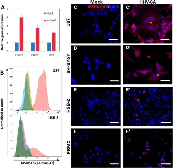
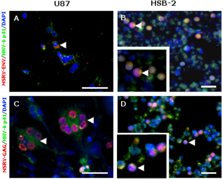
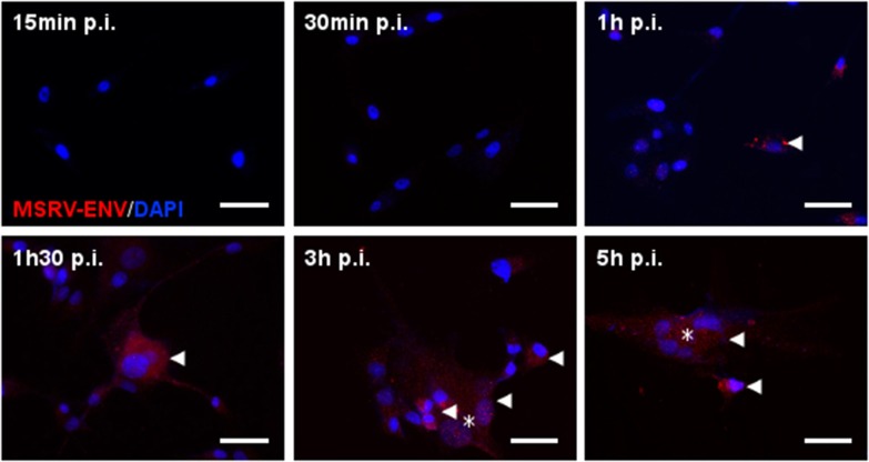
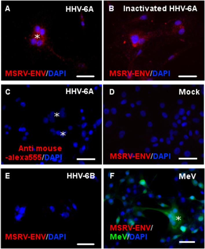
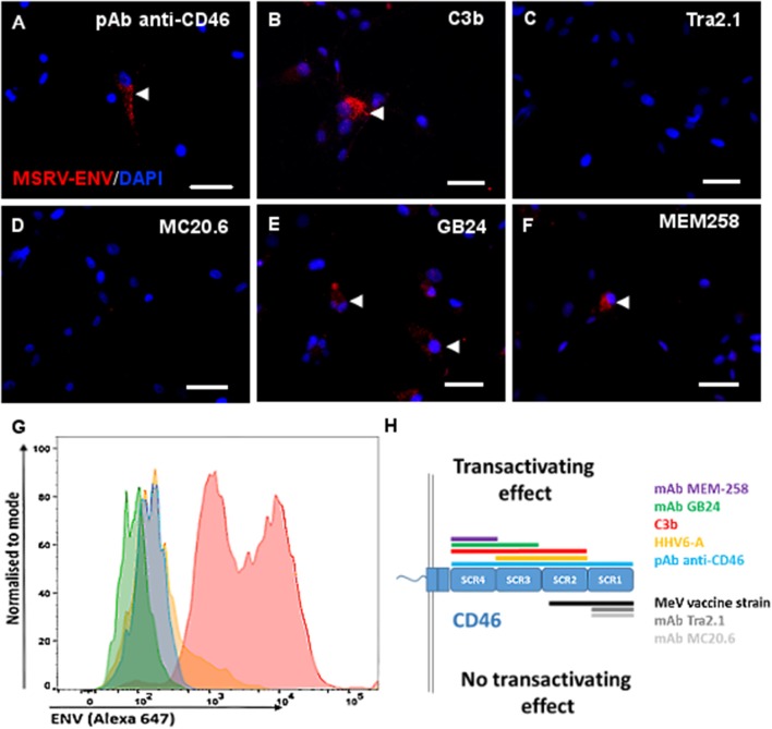
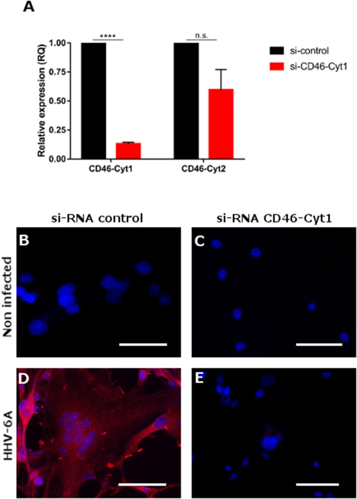
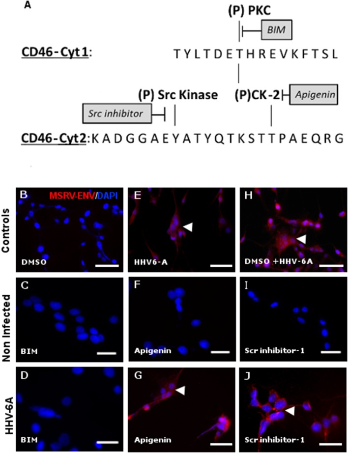
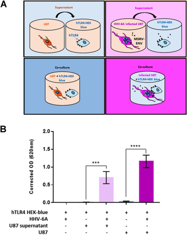
The aberrant expression of human endogenous retrovirus (HERV) elements of the HERV-W family has been associated with different diseases, including multiple sclerosis (MS). In particular, the expression of the envelope protein (Env) from the multiple sclerosis-associated retrovirus (MSRV), a member of HERV-W family and known for its potent proinflammatory activity, is repeatedly detected in the brain lesions and blood of MS patients. Furthermore, human herpesvirus 6 (HHV-6) infection has long been suspected to play a role in the pathogenesis of MS and neuroinflammation. We show here that both HHV-6A and stimulation of its receptor, transmembrane glycoprotein CD46, induce the expression of MSRV-Env. The engagement of extracellular domains SCR3 and SCR4 of CD46-Cyt1 isoform was required for MSRV-env transactivation, limiting thus the MSRV-Env induction to the CD46 ligands binding these domains, including C3b component of complement, specific monoclonal antibodies, and both infectious and UV-inactivated HHV-6A, but neither HHV-6B nor measles virus vaccine strain. Induction of MSRV-Env required CD46 Cyt-1 singling and was abolished by the inhibitors of protein kinase C. Finally, both membrane-expressed and secreted MSRV-Env trigger TLR4 signaling, displaying thus a proinflammatory potential, characteristic for this viral protein. These data expand the specter of HHV-6A effects in the modulation of the immune response and support the hypothesis that cross-talks between exogenous and endogenous viruses may contribute to inflammatory diseases and participate in neuroinflammation. Furthermore, they reveal a new function of CD46, known as an inhibitor of complement activation and receptor for several pathogens, in transactivation of HERV env genes, which may play an important role in the pathogenesis of inflammatory diseases.

Keywords: CD46; HHV-6A; MSRV; TLR4; human endogenous retrovirus; inflammation; multiple sclerosis.


Figures

Figure 1
Figure 1
HHV-6A infection induces expression of MSRV-Env in different cell types. (A) HSB2 cells, cord blood mononuclear cells (CBMC) and U87 cells were infected with HHV-6A at MOI 1 or incubated with the mock control for 24 h and MSRV env expression was analyzed by RT-qPCR. The values are expressed relatively to those in mock-infected cells and error bars represent SEM of 3 independent experiments. (B) Cytofluorometric analysis of U87 and HSB-2 cells infected or not with HHV-6A for 24 h at MOI 0.1 and stained by anti-MSRV-Env mAb, followed by anti-mouse Ig-Alexa 647 (green: non-infected cells + secondary Ab staining; orange: non-infected cells + complete staining; blue: infected cells + secondary Ab staining, pink: infected cells + complete staining). (C,C') U87, (D,D') SH-SY5Y, (E,E') HSB-2, and (F,F') peripheral blood mononuclear cells (PBMC) were either incubated with mock preparation (C–F) or infected at MOI 0.1 (C'–F') and analyzed 24 h later by immunofluorescence using anti-MSRV-Env mAb, followed by anti-mouse Ig-Alexa 555 (red staining). HHV-6A induced syncytia formation of adherent infected cells (C',D', *). DAPI (blue staining) was used to visualize cell nuclei (Bar = 50 μm). Data are representative of at least three independent experiments.
Figure 2
Figure 2
MSRV-ENV and MSRV-GAG were expressed in HHV-6A infected cells. (A,C) U87 and (B,D) HSB-2 cells were infected with HHV-6A during 24 h. Cells were fixed and stained for MSRV-ENV (GN_mAb_Env01) (A,B) or MSRV-GAG (GN_mAb_Gag06) (C,D) followed by anti-mouse-Alexa 555 (red staining). HHV-6 staining was revealed using biotinylated anti-HHV-6-p41 mAb, followed by streptavidin-FITC (green staining). HHV-6A infected cells expressing either MSRV-ENV or MSRV-GAG were pointed by arrowhead. DAPI (blue staining) was used to visualize cell nuclei. Bar = 50 μm. Bottom left frames (B,D): higher magnification of cell pointed by arrowhead.
Figure 3
Figure 3
Kinetics of HHV-6A-induced MSRV-Env production in U87 cells. U87 cells were infected with HHV-6A (MOI 1) and analyzed at indicated time points after infection by immunofluorescence using anti-MSRV-Env mAb, followed by anti-mouse-Alexa 555 (red staining, arrowhead). HHV-6A induced syncytia formation observed at 3 h p.i. (*). DAPI (blue staining) was used to visualize cell nuclei. Bar = 50 μm.
Figure 4
Figure 4
Both infectious and UV-inactivated HHV-6A, but neither HHV-6B nor Measles virus, could induce MSRV-Env expression. U87 cells were either infected or put in contact with UV-inactivated virus and observed 24 h later for the expression of MSRV-Env. Following viruses at MOI 1 were used: (A,C) HHV-6A, (B) UV-inactivated HHV-6A, (D) mock-infected control, (E) HHV-6B, and (F) recombinant measles virus (MeV) vaccine strain expressing EGFP (green staining, *syncytia formation). The expression of MSRV-Env was detected using anti-MSRV-Env mAb immunofluorescence analysis, followed by anti-mouse-Alexa 555 (red staining), including DAPI (blue staining) to visualize cell nuclei (bar = 50 μm). Alternatively, primary mAb was replaced with the isotype control (C).
Figure 5
Figure 5
Engagement of CD46-SCR3 and/or CD46-SCR4 trigers intracellular and cell surface MSRV-Env expression. Several CD46 ligands known to bind different CD46 SCR domains were used to stimulate U87 cells: (A,G) anti-CD46 rabbit polyclonal antibody, (B) C3b component of complement (50 mM), (C) Anti-SCR1 Tra2.1 (75 μg/ml), (D) anti-SCR1 MC20.6 (75 μg/ml), (E) anti-SCR3-4 GB24 (75 μg/ml), (F) anti-SCR4 MEM-258 (50μg/ml). The expression of MSRV-Env was detected using anti-MSRV-Env mAb-coupled to Alexa 555 by immunofluorescence analysis (red staining, arrowhead), including DAPI (blue staining) to visualize cell nuclei (bar = 50 μm) (A–F), or by cytofluorometry (G), using anti-MSRV-Env mAb followed with anti-mouse-Alexa 647 (blue: cells stimulated with anti-CD46 + secondary Ab staining, pink: cells stimulated with anti-CD46 followed by complete anti-MSRV-Env staining, green: cells stimulated with isotype control mAb + staining with secondary Ab; orange: cells stimulated with isotype control mAb followed by complete anti-MSRV-Env staining). Good cell viability was observed in all stimulation conditions. (H) Schematic representation of CD46 SCR domains recognized with utilized CD46 ligands, having a different effect on the induction of MSRV-Env.
Figure 6
Figure 6
Induction of MSRV-Env expression is dependent of CD46-Cyt1 isoforme. (A) CD46-Cyt1 and CD46-Cyt2 mRNA expression levels in si-CD46-Cyt1-treated U87 cells normalized to si-control treated cells, analyzed by RT-qPCR (relative expression). Error bars, mean ± SD of three experiments, statistical test used: nonparametric KS test (n.s. P > 0.1, ****P < 0.0001, vs. untreated condition) (B–E) MSRV-Env expression in U87 cells, treated with either si-RNA control (B,D) or and si-Cyt1 (C,E), and either left non-infected (B,C) or infected with HHV-6A (MOI 0.1) for 24 h (D,E). MSRV-Env expression was determined using anti-MSRV-Env mAb, followed by anti-mouse-Alexa 555 (red staining) cellular nuclei were visualized using DAPI (blue staining), bar = 50 μm.
Figure 7
Figure 7
Activation of MSRV-Env expression requires protein kinase C activity. (A) Schematic representation of putative phosphorylation sites within CD46-Cyt1 and Cyt2 and candidate kinases and their inhibitors (PKC: Protein Kinases C; CK-2: Casein Kinase 2). (B,E,H) Transactivation controls. (C,D) Effect of bisindolylmaleimide 5 μM (BIM) on HHV-6A-induced MSRV-Env expression; (F,G,I,J) absence of the effect of, Apigenin 40 μM and Scr-inhibitor 5 μM on MSRV-Env expression. The expression of MSRV-Env was analyzed by immuofluorescence, using anti-MSRV Env mAb, followed by anti-mouse-Alexa 555 (red staining, arrowhead) and representative images from 3 independent experiments were presented. DAPI (blue staining) was used to visualize cell nuclei, bar = 50 μm.
Figure 8
Figure 8
HHV-6A-induced MSRV-Env triggers human TLR4. (A) Schematic presentation of experimental conditions. HEK cells co-expressing human TLR4 (hTLR4) and TLR4 activation reporter gene (SEAP), were stimulated with either HHV-6A, supernatants from HHV-6A infected or non-infected U87 cells or co-cultured in presence of U87 cells previously infected with HHV-6A or not. hTLR4 activation was monitored colorimetrically using a SEAP reporter gene placed under the control of an NF-κB inducible promoter. (B) Data are expressed as corrected absorbance at 620 nm (absorbance obtained for HEK-blue + HHV-6A, mean: 0.27 ± 0.3, was subtracted from the absorbance obtained for other experimental conditions). Statistical analysis was performed using t-test on means ± SEM from three independent experiments (***P < 0.001, ****P < 0.0001, vs. uninfected conditions).

Similar articles

Cited by

References

    1. Bannert N, Kurth R. Retroelements and the human genome: new perspectives on an old relation. Proc Natl Acad Sci USA. (2004) 101(Suppl. 2):14572–9. 10.1073/pnas.0404838101 - DOI - PMC - PubMed
    1. Seifarth W, Frank O, Zeilfelder U, Spiess B, Greenwood AD, Hehlmann R, et al. . Comprehensive analysis of human endogenous retrovirus transcriptional activity in human tissues with a retrovirus-specific microarray. J Virol. (2005) 79:341–52. 10.1128/JVI.79.1.341-352.2005 - DOI - PMC - PubMed
    1. Frank O, Giehl M, Zheng C, Hehlmann R, Leib-Mösch C, Seifarth W. Human endogenous retrovirus expression profiles in samples from brains of patients with schizophrenia and bipolar disorders. J Virol. (2005) 79:10890–901. 10.1128/JVI.79.17.10890-10901.2005 - DOI - PMC - PubMed
    1. Perron H, Mekaoui L, Bernard C, Veas F, Stefas I, Leboyer M. Endogenous retrovirus type W GAG and envelope protein antigenemia in serum of schizophrenic patients. Biol Psychiatry (2008) 64:1019–23. 10.1016/j.biopsych.2008.06.028 - DOI - PubMed
    1. Balada E, Vilardell-Tarrés M, Ordi-Ros J. Implication of human endogenous retroviruses in the development of autoimmune diseases. Int Rev Immunol. (2010) 29:351–70. 10.3109/08830185.2010.485333 - DOI - PubMed

Publication types

MeSH terms