

Interleukin-33 Contributes Toward Loss of Tolerance by Promoting B-Cell-Activating Factor of the Tumor-Necrosis-Factor Family (BAFF)-Dependent Autoantibody Production
- PMID: 30574145
- PMCID: PMC6292404
- DOI: 10.3389/fimmu.2018.02871
Interleukin-33 Contributes Toward Loss of Tolerance by Promoting B-Cell-Activating Factor of the Tumor-Necrosis-Factor Family (BAFF)-Dependent Autoantibody Production
Abstract
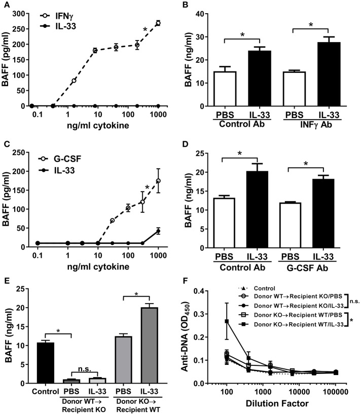
Breaking tolerance is a key event leading to autoimmunity, but the exact mechanisms responsible for this remain uncertain. Here we show that the alarmin IL-33 is able to drive the generation of autoantibodies through induction of the B cell survival factor BAFF. A temporary, short-term increase in IL-33 results in a primary (IgM) response to self-antigens. This transient DNA-specific autoantibody response was dependent on the induction of BAFF. Notably, radiation resistant cells and not myeloid cells, such as neutrophils or dendritic cells were the major source of BAFF and were critical in driving the autoantibody response. Chronic exposure to IL-33 elicited dramatic increases in BAFF levels and resulted in elevated numbers of B and T follicular helper cells as well as germinal center formation. We also observed class-switching from an IgM to an IgG DNA-specific autoantibody response. Collectively, the results provide novel insights into a potential mechanism for breaking immune-tolerance via IL-33-mediated induction of BAFF.
Keywords: B cell; BAFF; IL-33; autoantibodies; germinal center; immune tolerance; radiation resistant.
Figures


Similar articles
-
TACI deletion protects against progressive murine lupus nephritis induced by BAFF overexpression.Kidney Int. 2018 Oct;94(4):728-740. doi: 10.1016/j.kint.2018.03.012. Epub 2018 Jun 12. Kidney Int. 2018. PMID: 29907458 Free PMC article.
-
Myeloid cells, BAFF, and IFN-gamma establish an inflammatory loop that exacerbates autoimmunity in Lyn-deficient mice.J Exp Med. 2010 Aug 2;207(8):1757-73. doi: 10.1084/jem.20100086. Epub 2010 Jul 12. J Exp Med. 2010. PMID: 20624892 Free PMC article.
-
IL10 restrains autoreactive B cells in transgenic mice expressing inactive RAG1.Cell Immunol. 2018 Sep;331:110-120. doi: 10.1016/j.cellimm.2018.06.004. Epub 2018 Jun 13. Cell Immunol. 2018. PMID: 30017086 Free PMC article.
-
B cells flying solo.Immunol Cell Biol. 2008 Jan;86(1):40-6. doi: 10.1038/sj.icb.7100142. Immunol Cell Biol. 2008. PMID: 18172443 Review.
-
B-cell tolerance breakdown in Sjögren's syndrome: focus on BAFF.Autoimmun Rev. 2010 Jul;9(9):604-8. doi: 10.1016/j.autrev.2010.05.006. Epub 2010 May 8. Autoimmun Rev. 2010. PMID: 20457281 Review.
Cited by
-
Low Serum BAFF Concentration Is Associated with Response to TNF Inhibitors in Seropositive Patients with Rheumatoid Arthritis.J Clin Med. 2022 Sep 2;11(17):5207. doi: 10.3390/jcm11175207. J Clin Med. 2022. PMID: 36079136 Free PMC article.
-
Spatial Transcriptomics Resolve an Emphysema-Specific Lymphoid Follicle B Cell Signature in Chronic Obstructive Pulmonary Disease.Am J Respir Crit Care Med. 2024 Jan 1;209(1):48-58. doi: 10.1164/rccm.202303-0507LE. Am J Respir Crit Care Med. 2024. PMID: 37934672 Free PMC article.
-
Dysregulated B cell function and disease pathogenesis in systemic sclerosis.Front Immunol. 2023 Jan 16;13:999008. doi: 10.3389/fimmu.2022.999008. eCollection 2022. Front Immunol. 2023. PMID: 36726987 Free PMC article. Review.
-
High-throughput identification of autoantibodies that target the human exoproteome.Cell Rep Methods. 2022 Feb 28;2(2):100172. doi: 10.1016/j.crmeth.2022.100172. Epub 2022 Feb 17. Cell Rep Methods. 2022. PMID: 35360706 Free PMC article.
-
Diagnostic Evaluation Using Salivary Gland Ultrasonography in Primary Sjögren's Syndrome.J Clin Med. 2023 Mar 22;12(6):2428. doi: 10.3390/jcm12062428. J Clin Med. 2023. PMID: 36983428 Free PMC article.
References
MeSH terms
Substances
LinkOut - more resources
Full Text Sources
Medical
Molecular Biology Databases