. 2018 Dec 19:15:52.
doi: 10.1186/s12983-018-0293-6. eCollection 2018.

Also looking like Limulus? - retinula axons and visual neuropils of Amblypygi (whip spiders)

Affiliations

Also looking like Limulus? - retinula axons and visual neuropils of Amblypygi (whip spiders)

Tobias Lehmann et al. Front Zool. .

Abstract

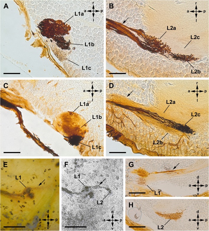
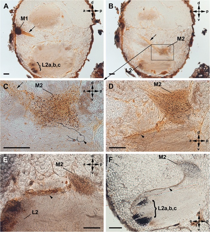
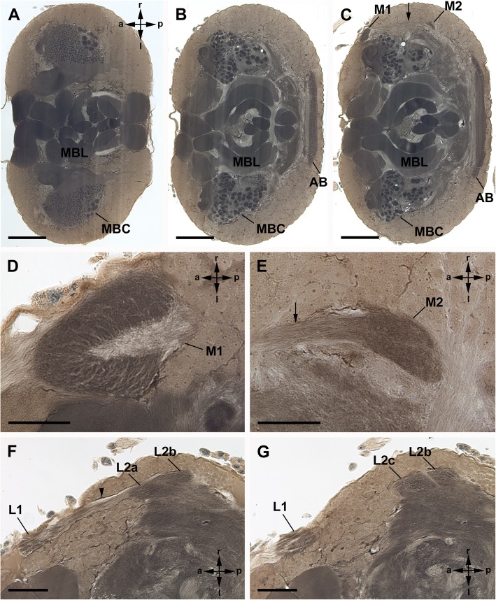
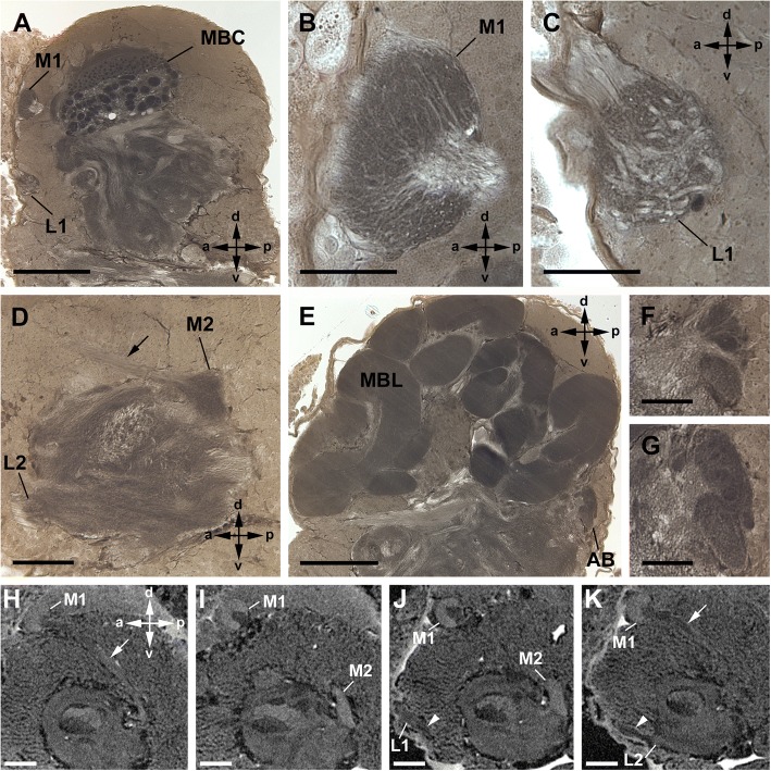
Background: Only a few studies have examined the visual systems of Amblypygi (whip spiders) until now. To get new insights suitable for phylogenetic analysis we studied the axonal trajectories and neuropil architecture of the visual systems of several whip spider species (Heterophrynus elaphus, Damon medius, Phrynus pseudoparvulus, and P. marginemaculatus) with different neuroanatomical techniques. The R-cell axon terminals were identified with Cobalt fills. To describe the morphology of the visual neuropils and of the protocerebrum generally we used Wigglesworth stains and μCT.

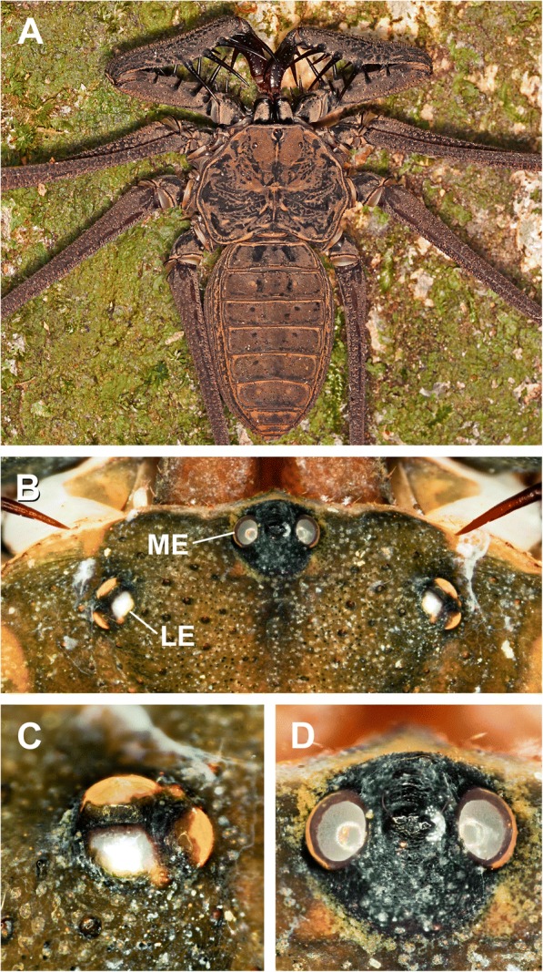
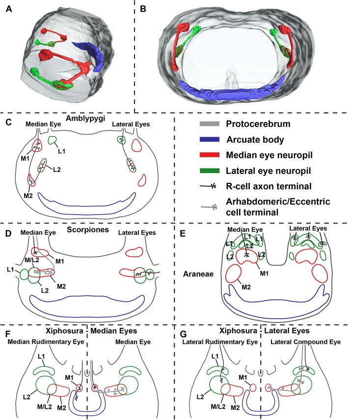
Results: The visual system of whip spiders comprises one pair of median and three pairs of lateral eyes. The R-cells of both eye types terminate each in a first and a second visual neuropil. Furthermore, a few R-cell fibres from the median eyes leave the second median eye visual neuropil and terminate in the second lateral eye neuropil. This means R-cell terminals from the lateral eyes and the median eyes overlap. Additionally, the arcuate body and the mushroom bodies are described.

Conclusions: A detailed comparison of our findings with previously studied chelicerate visual systems (i.e., Xiphosura, Scorpiones, Pseudoscorpiones, Opiliones, and Araneae) seem to support the idea of close evolutionary relationships between Xiphosura, Scorpiones, and Amblypygi.

Keywords: Arachnida; Central projections; Chelicerata; Phylogeny; Visual system.


Conflict of interest statement

Not applicable.Not applicable.The authors declare that they have no competing interests.Springer Nature remains neutral with regard to jurisdictional claims in published maps and institutional affiliations.

Figures

Fig. 1
Fig. 1
Median and lateral eyes in Amblypygi. Heterophrynus elaphus (a), Damon medius (b-d). a, Whip spider in natural habitat. b, Anterior part of the cephalothorax with one pair of median eyes and three pairs of lateral eyes. c, Detail of lateral eyes. d, Detail of median eyes
Fig. 2
Fig. 2
Cobalt fills via lateral eyes in Amblypygi. Damon medius; frontal sections (a-d); sagittal view (e-h). a, b, Cobalt fills via two lateral eyes, accordingly two subunits (subunit a, b) of first (a) and second (b) visual neuropils are stained. Note dense arrangement with varicosities of Cobalt-filled profiles. In the tract between the first and second neuropil (arrow) it seems, that the fibres cross. Bars 50 μm. c, d, Cobalt fills via one lateral eye (different eye than in a, b), accordingly different subunit (subunit c) of the first (c) and second (d) visual neuropil are stained than in a, b. Bars 50 μm. e-h, Comparison of whole mount, μCT, and sections of same specimen. Two lateral eyes are filled with cobalt, hence two subunits of the first lateral eye visual neuropil are stained. Dorsally the first and second visual neuropil is stained with cobalt, but ventrally cobalt stopped in the first visual neuropil, and the second neuropil remained unstained. In whole mount (e; bar 250 μm) only the first visual neuropil is definitely visible. In μCT reconstruction (f; bar 250 μm) the first and second neuropil is visible. Most details become apparent in sections (g, h; bars 100 μm). L1, first lateral eye neuropil; L2, second lateral eye neuropil; a-c subunits of L1 and L2; arrow, tract connecting L1 and L2
Fig. 3
Fig. 3
Cobalt fills via median eyes in Amblypygi. Phrynus marginemaculatus; sagittal sections (a-c); frontal sections (d-f). a, b, Two consecutive sections of the same specimen. a, First and second median eye visual neuropil filled with cobalt. Note dense arrangement of Cobalt-filled profiles in the first visual neuropil and just a few filled fibres running further to the second visual neuropil resulting in a diffuse staining of the neuropil. Both neuropils are connected via a tract (arrow). Bar 100 μm. b, First and second visual neuropil stained. Additionally, a thin tract connects the second median eye neuropil and the second lateral eye neuropil (arrowhead). A few cobalt filled R-cell axons run through this tract and terminate in the second lateral eye neuropil. Bar 100 μm. c, Same situation in a different specimen. Some cobalt filled R-cells from the median eye terminate in the second lateral eye visual neuropil. Bars 100 μm. d-f, Same situation in a third specimen and in frontal sections. Bars 100 μm. M1, first median eye visual neuropil; M2, second median eye visual neuropil; L1, first lateral eye neuropil; L2, second lateral eye neuropil; arrow, tract connecting M1 and M2; arrowhead thin tract connecting M2 and L2
Fig. 4
Fig. 4
Cobalt fills simultaneously via median and lateral eyes in Amblypygi. Phrynus pseudoparvulus; all eight eyes stained; sagittal sections. a-d, Three consecutive sections of the same specimen; c detail of b. Cobalt visible in (I) the first median eye visual neuropil, (II) the tract between first and second median eye neuropil (arrows), (III) the second median eye visual neuropil, (IV) a thin tract between the second median eye and second lateral eye visual neuropil (arrowheads), (V) the second lateral eye neuropil, (VI) the tract between the first and second median eye neuropil (not shown, but available on morph-d-base) and (VII) the first lateral eye neuropil (not shown, but available on morph-d-base). Note U-turn of a few fibres (arrowheads) from the second median eye neuropil to a thin tract connecting the second median and second lateral eye neuropil. Bars 50 μm. e, f, Different specimens showing details of the connection between the second median and second lateral eye neuropil via a thin tract (arrowhead). Bars 50 μm. M1, first median eye visual neuropil; M2, second median eye visual neuropil; L2, second lateral eye neuropil; arrow, tract connecting M1 and M2; a-c subunits of L2; arrowhead thin tract connecting M2 and L2
Fig. 5
Fig. 5
General anatomy of visual neuropils and protocerebrum in Amblypygi, frontal sections. Heterophrynus elaphus; Wigglesworth stains. a-c, Three sections from dorsal (a), central (b) and more ventral (c) part of the protocerebrum showing large-scale spread of the mushroom bodies, especially of the mushroom body lobes. Note the relatively small arcuate body. Bars 250 μm. d, e, Detail of the first and second median eye visual neuropil. Bars 50 μm. f, g, Detail of the first and second lateral eye visual neuropil. Note subdivision of the second visual neuropil into three subunits. Bars 100 μm. AB, arcuate body; MBC, mushroom body calyces; MBL, mushroom body lobes; M1, first median eye visual neuropil; M2, second median eye visual neuropil; L1, first lateral eye neuropil; L2, second lateral eye neuropil; arrow, tract connecting M1 and M2; a-c subunits of L2; arrowhead tract connecting L1 and L2
Fig. 6
Fig. 6
General anatomy of visual neuropils and protocerebrum in Amblypygi, sagittal sections. Heterophrynus elaphus (a-g) and Damon medius (h-k); Wigglesworth stains (a-g) and μCT (h-k, additional raw data on morph-D-base). a, Dorsal to the first median eye visual neuropil lies the first lateral eye neuropil. Bar 250 μm. b, c, Detail of the first median and lateral eye visual neuropil. Bars 50 μm. d, Section showing a tract connecting the first and second median eye neuropil, the second median eye neuropil, and the second lateral eye neuropil. Bar 100 μm. E, Mid-sagittal section showing the mushroom body lobes filling almost the entire protocerebrum. Note the relatively small arcuate body. Bar 250 μm. f, g, Detail of the arcuate body in parasagittal (f) and midsagittal (g) region. Bars 50 μm. h, i, μCT reconstruction showing the first and second median eye visual neuropil and a tract (arrow) connecting these two neuropils. Bars 250 μm. j, k, μCT reconstruction showing the first and second lateral eye visual neuropil and a tract (arrow) connecting these two neuropils. Bars 250 μm. AB, arcuate body; MBC, mushroom body calyces; MBL, mushroom body lobes; M1, first median eye visual neuropil; M2, second median eye visual neuropil; L1, first lateral eye neuropil; L2, second lateral eye neuropil; arrow, tract connecting M1 and M2; arrowhead tract connecting L1 and L2
Fig. 7
Fig. 7
Comparison of median and lateral eye visual systems in (c) Amblypygi, (d) Scorpiones, (e) Araneae, and (f, g) Xiphosura. a, b, 3D serial reconstruction of the visual system of whip spider Heterophrynus elaphus. c, Amblypygi (Damon medius, Heterophrynus elaphus, Phrynus spp.), this study. d, Scorpiones (Euscorpius spp., Androctonus australis), after Lehmann, et al. [33] and Fleissner [40]. e, Araneae (Cupiennius salei), after Strausfeld, et al. [28], Strausfeld, et al. [29]. f, g, Xiphosura (Limulus polyphemus), after Lehmann, et al. [33], Calman, et al. [38], Chamberlain, et al. [39]. L1, first lateral eye visual neuropil; L2, second lateral eye visual neuropil; M1, first median eye visual neuropil; M2, second median eye visual neuropil; M/L2, region were M1 and L1 overlap

Similar articles

Cited by

References

    1. Whip spiders of the World, version 1.0. Western Australian Museum, Perth. http://museum.wa.gov.au/catalogues-beta/whip-spiders
    1. Weygoldt P. Whip spiders (Chelicerata, Amblypygi). Stenstrup: Apollo Books; 2000.
    1. Dunlop JA. Systematics of the coal measures whip spiders (Arachnida: Amblypygi) Zool Anz. 2018;273:14–22. doi: 10.1016/j.jcz.2017.11.004. - DOI
    1. Dunlop JA. Geological history and phylogeny of Chelicerata. Arthropod Struct Dev. 2010;39:124–142. doi: 10.1016/j.asd.2010.01.003. - DOI - PubMed
    1. Bingman VP, Graving JM, Hebets EA, Wiegmann DD: Importance of the antenniform legs, but not vision, for homing by the neotropical whip spider, Paraphrynus laevifrons J Exp Biol. 2016;jeb:149823. - PubMed

LinkOut - more resources