

Two Uveal Melanomas in One Eye: A Choroidal Nevus Giving Rise to a Melanoma in an Eye with a Separate Large Choroidal Melanoma
- PMID: 30574486
- PMCID: PMC6288664
- DOI: 10.1159/000486682
Two Uveal Melanomas in One Eye: A Choroidal Nevus Giving Rise to a Melanoma in an Eye with a Separate Large Choroidal Melanoma
Abstract
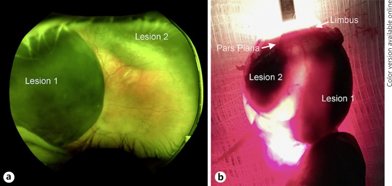
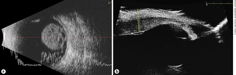
Multifocal uveal melanomas are extremely rare. In this case report, we describe a patient with 2 independent uveal melanomas in the same eye. A 52-year-old woman presented with a large choroidal melanoma and a smaller ciliary body mass, clinically thought to be a nevus, in her left eye. Enucleated specimen showed 2 primary lesions that were anatomically separate. Lesion 1 was a melanoma and lesion 2 was a melanoma arising centrally from a nevus. Both lesions harbored GNAQ mutations. This patient had no family history of uveal melanomas or signs of ocular melanocytosis and was negative for the BAP1 mutation. This case demonstrates how multifocal uveal melanomas can arise in patients who lack genetic predisposition for the disease. Furthermore, this case is one of the few that have shown, histopathologically, a small focus of malignant cells developing from a benign population within a nevus, which highlights the importance of closely monitoring nevi for signs of malignancy.
Keywords: Choroidal melanoma; Choroidal nevus; Histopathology; Multifocal melanoma; Multiple melanoma; Uveal melanoma.
Figures

References
-
- Nissen EJ, Kalirai H, Damato B, Heimann H, Coupland SE. Two distinct uveal melanomas in the same eye. JAMA Ophthalmol. 2015;133:1094–1096. - PubMed
-
- Dithmar S, Volcker HE, Grossniklaus HE. Multifocal intraocular malignant melanoma: report of two cases and review of the literature. Ophthalmology. 1999;106:1345–1348. - PubMed
-
- Hadden PW, Damato BE. Consecutive choroidal melanoma in the same eye of a patient. Am J Ophthalmol. 2003;135:728–729. - PubMed
-
- Blumenthal EZ, Pe'er J. Multifocal choroidal malignant melanoma: at least 3 melanomas in one eye. Arch Ophthalmol. 1999;117:255–258. - PubMed
Grants and funding
LinkOut - more resources
Full Text Sources
