. 2019 Feb 12;58(6):621-632.
doi: 10.1021/acs.biochem.8b01045. Epub 2019 Jan 10.

UBC9 Mutant Reveals the Impact of Protein Dynamics on Substrate Selectivity and SUMO Chain Linkages

UBC9 Mutant Reveals the Impact of Protein Dynamics on Substrate Selectivity and SUMO Chain Linkages

Christine M Wright et al. Biochemistry. .

Abstract

SUMO, a conserved ubiquitin-like protein, is conjugated to a multitude of cellular proteins to maintain genomic integrity and resist genotoxic stress. Studies of the SUMO E2 conjugating enzyme mutant, UBC9P123L, suggested that altered substrate specificity enhances cell sensitivity to DNA damaging agents. Using nuclear magnetic resonance chemical shift studies, we confirm that the mutation does not alter the core globular fold of UBC9, while 15N relaxation measurements demonstrate mutant-induced stabilization of distinct chemical states in residues near the active site cysteine and substrate recognition motifs. We further demonstrate that the P123L substitution induces a switch from the preferential addition of SUMO to lysine residues in unstructured sites to acceptor lysines embedded in secondary structures, thereby also inducing alterations in SUMO chain linkages. Our results provide new insights regarding the impact that structural dynamics of UBC9 have on substrate selection and specifically SUMO chain formation. These findings highlight the potential contribution of nonconsensus SUMO targets and/or alternative SUMO chain linkages on DNA damage response and chemotherapeutic sensitivity.


Figures
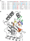
Figure 1:
Figure 1:. SUMO consensus interactions with UBC9.
(A) Sequence alignment of N-terminal residues from yeast SMT3, and human SUMO−1, SUMO−2, SUMO−3, SUMO−4, and SUMO−5. Consensus lysine residues are highlighted in red while non-consensus lysines are highlighted in cyan. (B) Ribbon presentation of UBC9 depicting location of P123 (yellow) and residues that are critical for SUMO transfer. Pro123 is located in the α2-α3 loop, and in coordination with Leu94, Ser95, Glu122, and Asn124 (orange), forms a series of stabilizing hydrogen bonds with the SUMO carboxy-terminal di-glycine motif [7]. Further along the same loop, residue Asp127, in concert with Asn85 and Tyr87 (blue), has been shown to catalyze SUMO conjugation by manipulating the pKa of the acceptor lysine residue [4, 7]. The carboxy-terminus of the α2-α3 loop contains residues Pro128, Gln130, and Ala131 (green), which make Van der Waals contacts with the conserved hydrophobic residue in the consensus SUMO acceptor site [ΨKx(D/E)] [7, 25]. Hydrogen bonding interactions with the consensus acidic D/E residues (in the acceptor site) is mediated by interactions with residues Lys74, Ser89, and Thr91 (red) [7], which reside in the loop that connects β4 with the 310 helix and also contains the active site cysteine, C93 (yellow). PDB: 1A3S
Figure 2:
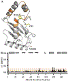
Figure 2:. Spectral response to P123L mutation.
(A) Overlay of 2D [15N, 1H]−HSQC spectra of wild-type (black) and P123L mutant (red) UBC9. (B) Se-quence mapping of weighted [ 15N, 1H] chemical shift perturbation resulting from mutation. Thresholds corresponding to 3 (0.05 ppm), 6 (0.1 ppm), and 9 (0.15 ppm) standard deviations are shown marked by yellow, orange or red dotted lines, respectively. Unassigned residues in the mutant spectra are denoted by negative cyan bars and the mutation site is identified by a negative gold bar. (C) Ribbon representation of the human UBC9 structure with locations of unassigned, moderately shifted and significantly shifted residues colored as described in (B). PDB: 1A3S
Figure 3:
Figure 3:. Spectral response to P123A mutation.
(A) Ribbon representation of the human UBC9 structure with locations of moderately shifted and significantly shifted residues colored as outlined below. (B) Sequence mapping of weighted [15N,1H] chemical shift perturbation resulting from P123A mutation. Threshold corresponding to 0.05 ppm and 0.1 ppm are marked by yellow or orange dotted lines, respectively. PDB: 1A3S
Figure 4:
Figure 4:. P123L retains secondary structured elements.
Plots of predicted alpha helical (positive, green) and beta strand (negative, blue) secondary structure derived from Cα and Cβ chemical shift values for (A) wild-type UBC9, (B) P123A, and (C) P123L. Unassigned residues in P123L are identified by red dashes. (D) Overlay of (A) and (C) showing a strong retention of secondary structure in the P123L mutant.
Figure 5:
Figure 5:. P123L mutation does not increase backbone flexibility.
Sequence plot of 15N−{1H} NOE intensities for (A) WT UBC9, (B) P123A, and (C) P123L. Unassigned residues and weak peaks (cyan dashes), split peaks (magenta circles), and a dashed line indicating significance thresholds (0.6; 0.0) are identified. (D) Mapping of the data illustrated in (C) on the structure of UBC9. Side chains of the hydrophobic residues F82, Y137, and Y144 are shown in grey. (E) Comparison of peak intensities obtained with (NONOE) and without (NOE) proton saturation, are presented in black and red, respectively. PDB: 1A3S
Figure 6:
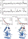
Figure 6:. Transverse relaxation rates (R2) indicate P123L stabilizes ns-μs chemical states.
Sequence plots of R2 values for (A) UBC9 and (B) P123L. Open circles mapped for residues with large and uncertain R2 relaxa-tion rates. Representative 15N−R2 relaxation data for (C) UBC9 and (D) P123L. The solid lines are single-exponential best fits. Cartoon plots of (E) UBC9 and (F) P123L, respectively mapping R2 rates on the structures. Color is based on R2 rates. Blue represents residues below the dashed threshold, indicative of free exchange, orange residues indicate a moderate increase in R2 rates, and red indicates residues with open circles in (A), (B) with large R2 relation rates. Missing assignments and residues K59, H83, and S89 with low R2 spectral intensity are colored in light blue. PDB: 1A3S
Figure 7:
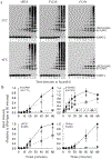
Figure 7:. P123L induces a switch in SUMO−2 chain linkage formation.
(A) Purified human E1, UBC9 (wild-type, P123A, or P123L) and SUMO−2 (molar ratio of 1:2.5:10, respectively) were incubated at 30°C, 37°C, or 42°C for 2 hours. 3X reactions contained 3-fold higher levels of P123L. Samples were immunoblotted with anti-SUMO−2 antibodies. A darker exposure of the portion of the gel boxed in red is shown below. (B) As in A, except as indicated wild-type or mutant SUMO−2 were included, and reactions sampled at varying times. Representative images of n=3 experiments.
Figure 8:
Figure 8:. Decreased global SUMO conjugation and N-terminal SUMO chain linkage formation by P123L.
(A) As in Figure 7, SUMOylation reactions were sampled at the indicated times, and immunoblotted with SUMO−2 antibodies. * position of free SUMO−2, ** indicates di-SUMO bands. (B) Band pixel densities (n=3 independent experiments) from 0–60 minutes, at 42°C, were quantified, with values plotted relative to those obtained with wild-type UBC9 at 60 minutes.

Similar articles

Cited by

References

    1. Bergink S; Jentsch S(2009) Principles of ubiquitin and SUMO modifications in DNA repair. Nature 458(7237), 461–7. - PubMed
    1. Seeler J-S; Dejean A(2017) SUMO and the robustness of cancer. Nat Rev Cancer 17(3), 184–197 - PubMed
    1. Flotho A; Melchior F(2013) Sumoylation: A Regulatory Protein Modification in Health and Disease. Annu Rev Biochem 82 (1), 357–385. - PubMed
    1. Capili AD; Lima CD(2007) Taking it step by step: mechanistic insights from structural studies of ubiquitin/ubiquitin-like protein modification pathways. Current Opinion in Structural Biology 17(6), 726–735. - PMC - PubMed
    1. Reverter D; Lima CD(2005) Insights into E3 ligase activity revealed by a SUMO-RanGAP1-Ubc9-Nup358 complex. Nature 435 (7042), 687–92. - PMC - PubMed

Publication types

MeSH terms