

Chlorophagy is ATG gene-dependent microautophagy process
- PMID: 30574829
- PMCID: PMC6351093
- DOI: 10.1080/15592324.2018.1558679
Chlorophagy is ATG gene-dependent microautophagy process
Abstract
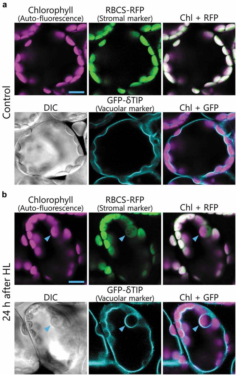
Autophagy delivers cytosolic components to lysosomes and the vacuole for degradation. This pathway prevents starvation through bulk degradation and recycling of cytoplasmic components, and maintains cellular homeostasis through selective elimination of damaged proteins and organelles. Autophagic delivery processes are categorized into three types: macroautophagy, microautophagy, and chaperone-mediated autophagy. During macroautophagy, nascent, double membrane-bound vesicles termed autophagosomes sequester a portion of cytoplasm and deliver it to the vacuole/lysosomes. Molecular genetic studies in budding yeasts have identified a set of AUTOPHAGY (ATG) genes required for autophagosome formation. Although microautophagy involves the direct lysosomal/vacuolar engulfment and incorporation of a target into the lumen rather than the formation of autophagosomes, the membrane dynamics and possible roles of ATGs during microautophagy are under investigation. Our recent study revealed an ATG-dependent microautophagy process in plants, during which chloroplasts damaged by high visible light (HL) are selectively eliminated. Here, we discuss the membrane dynamics of the plant microautophagy that enables the transport of whole chloroplasts into the vacuole.
Keywords: Autophagy; chlorophagy; chloroplast; microautophagy; photodamage.
Figures

Similar articles
-
Lysosomal microautophagy: an emerging dimension in mammalian autophagy.Trends Cell Biol. 2024 Jul;34(7):606-616. doi: 10.1016/j.tcb.2023.11.005. Epub 2023 Dec 15. Trends Cell Biol. 2024. PMID: 38104013 Review.
-
The core autophagy machinery is not required for chloroplast singlet oxygen-mediated cell death in the Arabidopsis thaliana plastid ferrochelatase two mutant.BMC Plant Biol. 2021 Jul 19;21(1):342. doi: 10.1186/s12870-021-03119-x. BMC Plant Biol. 2021. PMID: 34281507 Free PMC article.
-
Entire Photodamaged Chloroplasts Are Transported to the Central Vacuole by Autophagy.Plant Cell. 2017 Feb;29(2):377-394. doi: 10.1105/tpc.16.00637. Epub 2017 Jan 25. Plant Cell. 2017. PMID: 28123106 Free PMC article.
-
Partial or entire: Distinct responses of two types of chloroplast autophagy.Plant Signal Behav. 2017 Nov 2;12(11):e1393137. doi: 10.1080/15592324.2017.1393137. Epub 2017 Oct 17. Plant Signal Behav. 2017. PMID: 29040052 Free PMC article.
-
Microautophagy in Plants: Consideration of Its Molecular Mechanism.Cells. 2020 Apr 4;9(4):887. doi: 10.3390/cells9040887. Cells. 2020. PMID: 32260410 Free PMC article. Review.
Cited by
-
Plants' Response to Abiotic Stress: Mechanisms and Strategies.Int J Mol Sci. 2023 Jun 30;24(13):10915. doi: 10.3390/ijms241310915. Int J Mol Sci. 2023. PMID: 37446089 Free PMC article. Review.
-
The relationship between autophagy and respiratory viruses.Arch Microbiol. 2024 Mar 4;206(4):136. doi: 10.1007/s00203-024-03838-3. Arch Microbiol. 2024. PMID: 38436746 Review.
-
Diallyl disulfide and diallyl trisulfide in garlic as novel therapeutic agents to overcome drug resistance in breast cancer.J Pharm Anal. 2022 Apr;12(2):221-231. doi: 10.1016/j.jpha.2021.11.004. Epub 2021 Nov 10. J Pharm Anal. 2022. PMID: 35582397 Free PMC article. Review.
-
Autophagy: a game changer for plant development and crop improvement.Planta. 2022 Oct 28;256(6):103. doi: 10.1007/s00425-022-04004-z. Planta. 2022. PMID: 36307739 Review.
-
Genome-Wide Identification of ATG Gene Family Members in Fagopyrum tataricum and Their Expression during Stress Responses.Int J Mol Sci. 2022 Nov 27;23(23):14845. doi: 10.3390/ijms232314845. Int J Mol Sci. 2022. PMID: 36499172 Free PMC article.
References
Publication types
MeSH terms
LinkOut - more resources
Full Text Sources