

Pythium keratitis in South India: Incidence, clinical profile, management, and treatment recommendation
- PMID: 30574890
- PMCID: PMC6324135
- DOI: 10.4103/ijo.IJO_445_18
Pythium keratitis in South India: Incidence, clinical profile, management, and treatment recommendation
Abstract
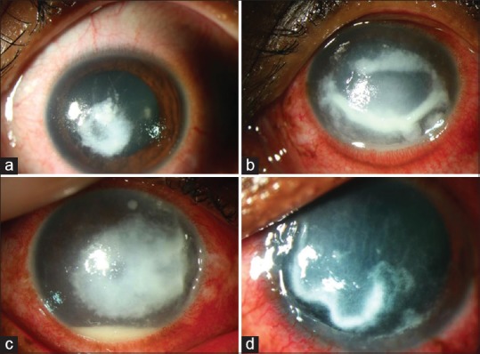
Purpose: To study the demographic profile, clinical features, treatment outcome, and ocular morbidity of microbiologically proven Pythium keratitis in South India.
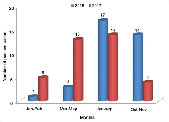
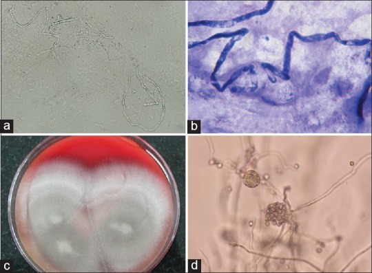
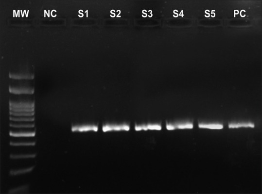
Methods: A retrospective analysis of clinical records of microbiologically proven Pythium keratitis at a tertiary eye care referral center in South India from January 2016 to November 2017 was performed. Demographic details, predisposing risk factors, microbiological investigations, clinical course, and visual outcome were analyzed.
Results: Seventy-one patients with microbiologically proven Pythium keratitis were identified. The mean age was 44(±18.2) years with an increase in male preponderance and 50% were farmers. Duration of delay at time of presentation to the hospital was a mean of 14(±7.2) days. The visual acuity at baseline ranged from 6/6 to no light perception (median 2.1 logMAR). A combination of 5% natamycin and 1% voriconazole was given to 42% patients, and natamycin alone was given to 39.4% patients. 1% itraconazole eye drops alone was initiated in 7 (10%) patients and 3 among this group responded. Therapeutic keratoplasty (TPK) was performed in 48 (67.6%) patients. None of the primary grafts remained clear after a period of 1 month. Twenty-six eyes (54.2%) had graft reinfection and all these eyes either developed anterior staphyloma (4) or were eviscerated (3) and 13 eyes became phthisical. The remaining 22 patients who had TPK resulted in failed graft. Among these, re-grafts were performed in 6 patients, of which 5 were doing well at the last follow-up.
Conclusion: We report a large series of patients with Pythium keratitis. Promoting early and differential diagnosis, awareness of clinicians and specific treatment options are needed for this devastating corneal disease.
Keywords: DNA sequencing; ITS; Pythium insidiosum; keratitis; therapeutic penetrating keratoplasty.
Conflict of interest statement
None
Figures
Comment in
-
Commentary: Pythium insidiosum keratitis.Indian J Ophthalmol. 2019 Jan;67(1):47-48. doi: 10.4103/ijo.IJO_1491_18. Indian J Ophthalmol. 2019. PMID: 30574891 Free PMC article. No abstract available.
-
Clinical differentiation of keratitis due to fungus and Pythium: A photographic survey.Indian J Ophthalmol. 2023 Feb;71(2):510-514. doi: 10.4103/ijo.IJO_913_22. Indian J Ophthalmol. 2023. PMID: 36727350 Free PMC article.
References
-
- Virgile R, Perry HD, Pardanani B, Szabo K, Rahn EK, Stone J, et al. Human infectious corneal ulcer caused by Pythium insidiosum. Cornea. 1993;12:81–3. - PubMed
-
- Thanathanee O, Enkvetchakul O, Rangsin R, Waraasawapati S, Samerpitak K, Suwan-Apichon O, et al. Outbreak of Pythium keratitis during rainy season: A case series. Cornea. 2013;32:199–204. - PubMed
-
- Lekhanont K, Chuckpaiwong V, Chongtrakool P, Aroonroch R, Vongthongsri A. Pythium insidiosum keratitis in contact lens wear: A case report. Cornea. 2009;28:1173–7. - PubMed
-
- Badenoch PR, Mills RA, Chang JH, Sadlon TA, Klebe S, Coster DJ, et al. Pythium insidiosum keratitis in an Australian child. Clin Exp Ophthalmol. 2009;37:806–9. - PubMed
-
- Tanhehco TY, Stacy RC, Mendoza L, Durand ML, Jakobiec FA, Colby KA, et al. Pythium insidiosum keratitis in Israel. Eye Contact Lens. 2011;37:96–8. - PubMed
MeSH terms
Substances
LinkOut - more resources
Full Text Sources
