

Deep learning in radiology: An overview of the concepts and a survey of the state of the art with focus on MRI
- PMID: 30575178
- PMCID: PMC6483404
- DOI: 10.1002/jmri.26534
Deep learning in radiology: An overview of the concepts and a survey of the state of the art with focus on MRI
Abstract
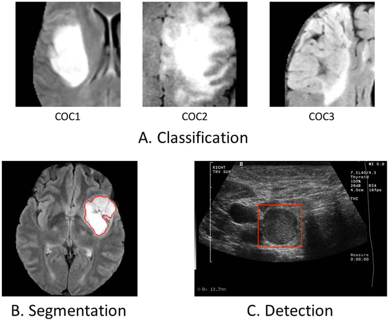
Deep learning is a branch of artificial intelligence where networks of simple interconnected units are used to extract patterns from data in order to solve complex problems. Deep-learning algorithms have shown groundbreaking performance in a variety of sophisticated tasks, especially those related to images. They have often matched or exceeded human performance. Since the medical field of radiology mainly relies on extracting useful information from images, it is a very natural application area for deep learning, and research in this area has rapidly grown in recent years. In this article, we discuss the general context of radiology and opportunities for application of deep-learning algorithms. We also introduce basic concepts of deep learning, including convolutional neural networks. Then, we present a survey of the research in deep learning applied to radiology. We organize the studies by the types of specific tasks that they attempt to solve and review a broad range of deep-learning algorithms being utilized. Finally, we briefly discuss opportunities and challenges for incorporating deep learning in the radiology practice of the future. Level of Evidence: 3 Technical Efficacy: Stage 1 J. Magn. Reson. Imaging 2019;49:939-954.
Keywords: artificial intelligence; convolutional neural networks; deep learning; machine learning; medical imaging; radiology.
© 2018 International Society for Magnetic Resonance in Medicine.
Figures
References
-
- Liu W, Wang Z, Liu X, Zeng N, Liu Y, Alsaadi FE: A survey of deep neural network architectures and their applications. Neurocomputing 2017; 234:11–26.
-
- Krizhevsky A, Sutskever I, Hinton GE: Imagenet classification with deep convolutional neural networks. In Adv Neural Inf Process Syst; 2012:1097–1105.
-
- Dodge S, Karam L: A Study and Comparison of Human and Deep Learning Recognition Performance Under Visual Distortions. arXiv Prepr arXiv170502498 2017.
-
- Rajpurkar P, Irvin J, Zhu K, et al. : CheXNet: Radiologist-Level Pneumonia Detection on Chest X-Rays with Deep Learning. arXiv Prepr arXiv171105225 2017.
-
- He K, Zhang X, Ren S, Sun J: Delving deep into rectifiers: Surpassing human-level performance on imagenet classification. In Proc IEEE Int Conf Comput Vis; 2015:1026–1034.
Publication types
MeSH terms
Grants and funding
LinkOut - more resources
Full Text Sources
Other Literature Sources
Medical
