Practice Guideline
. 2019 Jan;26(1):9-42.
doi: 10.1002/jhbp.596. Epub 2019 Jan 18.

Clinical practice guidelines for IgG4-related sclerosing cholangitis

Affiliations
Practice Guideline

Clinical practice guidelines for IgG4-related sclerosing cholangitis

Terumi Kamisawa et al. J Hepatobiliary Pancreat Sci. 2019 Jan.

Abstract

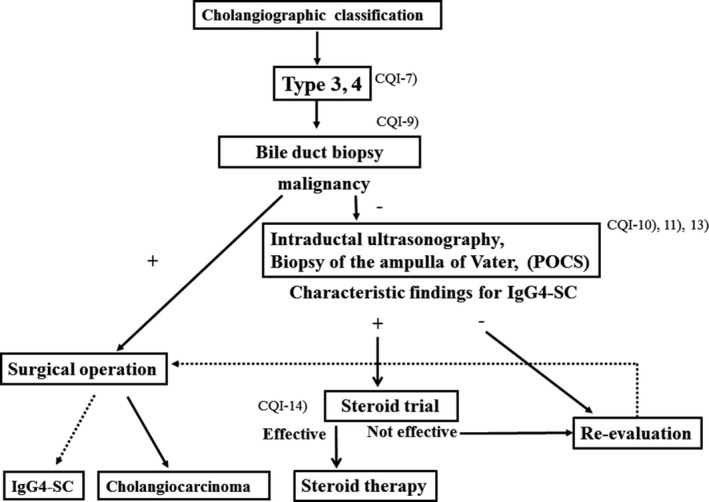
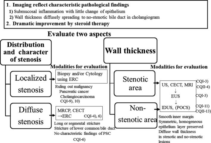
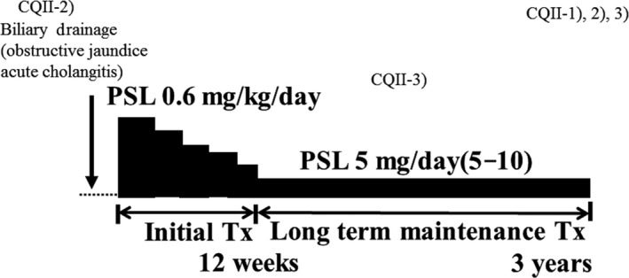
IgG4-related sclerosing cholangitis (IgG4-SC) is a distinct type of cholangitis frequently associated with autoimmune pancreatitis and currently recognized as a biliary manifestation of IgG4-related disease. Although clinical diagnostic criteria of IgG4-SC were established in 2012, differential diagnosis from primary sclerosing cholangitis and cholangiocarcinoma is sometimes difficult. Furthermore, no practical guidelines for IgG4-SC are available. Because the evidence level of most articles retrieved through searching the PubMed, Cochrane Library, and Igaku Chuo Zasshi databases was below C based on the systematic review evaluation system of clinical practice guidelines MINDS 2014, we developed consensus guidelines using the modified Delphi approach. Three committees (a guideline creating committee, an expert panelist committee for rating statements according to the modified Delphi method, and an evaluating committee) were organized. Eighteen clinical questions (CQs) with clinical statements were developed regarding diagnosis (14 CQs) and treatment (4 CQs). Recommendation levels for clinical statements were set using the modified Delphi approach. The guidelines explain methods for accurate diagnosis, and safe and appropriate treatment of IgG4-SC.

Keywords: Bile duct; Guideline; IgG4; Sclerosing cholangitis; Steroid.


Conflict of interest statement

None declared.

Figures

Figure 1
Figure 1
Cholangiographic classification of IgG4‐SC referred from Reference 18
Figure 2
Figure 2
Histological findings of transmural inflammatory cell infiltration and fibrosis in the bile duct of IgG4‐SC (arrow). (Scale: 100 μm)
Figure 3
Figure 3
Histological findings of storiform fibrosis in the bile duct of IgG4‐SC. (Scale: 100 μm)
Figure 4
Figure 4
Histological findings of obliterative phlebitis (arrows) in the bile duct of IgG4‐SC. (Scale: 100 μm)
Figure 5
Figure 5
Immunohistochemical findings of infiltration of numerous IgG4‐positive cells in the bile duct of IgG4‐SC. IgG4‐immunostaining. (Scale: 100 μm)
Figure 6
Figure 6
Algorithm for diagnosis and treatment of IgG4‐SC referred from Reference 18
Figure 7
Figure 7
Algorithm for diagnosis and treatment of IgG4‐SC referred from Reference 18
Figure 8
Figure 8
Algorithm for diagnosis and treatment of IgG4‐SC referred from Reference 18
Figure 9
Figure 9
Algorithm for diagnosis and treatment of IgG4‐SC referred from Reference 18
Figure 10
Figure 10
Summary of imaging findings suggestive of IgG4‐SC
Figure 11
Figure 11
Schematic illustration of treatment of IgG4‐SC
Figure 12
Figure 12
Ultrasonographic image of IgG4‐SC. Ultrasonography detects marked wall thickening in the extrahepatic bile duct (arrows)
Figure 13
Figure 13
EUS image of IgG4‐SC. EUS shows a homogeneous, high‐low‐high echo of the bile duct wall thickening (arrows)
Figure 14
Figure 14
CT image of IgG4‐SC. Contrast‐enhanced CT (delayed image) demonstrates concentric wall thickening of the hilar bile duct (arrows)
Figure 15
Figure 15
CT and MRCP images of IgG4‐SC. (a) In CT, the hilar bile duct shows marked thickening (arrow). (b) MRCP demonstrates bile duct strictures in the hilar bile duct (arrows)
Figure 16
Figure 16
ERCP image of a patient with AIP in which the lesion was limited to the pancreatic head. Narrowing of the main pancreatic duct in the head (arrows) and strictures of the lower bile ducts are recognized
Figure 17
Figure 17
IgG4‐related dacryoadenitis. Swelling of right and left lacrimal glands (right > left) is recognized (arrows)
Figure 18
Figure 18
CT image of IgG4‐related retroperitoneal fibrosis. Soft‐tissue mass surrounding the abdominal aorta is recognized (arrow)
Figure 19
Figure 19
Cholangiographic classification of IgG4‐SC and the differential diagnosis of IgG4‐SC referred from Reference 3
Figure 20
Figure 20
Schematic illustration comparing the cholangiographic findings in PSC and IgG4‐SC referred from Reference 3
Figure 21
Figure 21
Cholangiogram of PSC showing (a) band‐like strictures and (b) beaded appearance (arrows)
Figure 22
Figure 22
Cholangiogram of PSC showing (a) pruned‐tree appearance and (b) diverticulum‐like outpouching (arrows)
Figure 23
Figure 23
Cholangiopancreatogram of type 1 IgG4‐SC showing the lower bile duct stricture and irregular narrowing of the main pancreatic duct (arrows)
Figure 24
Figure 24
Cholangiogram of (a) type 2a IgG4‐SC showing dilatation after a confluent stricture (arrow) and (b) type 2b IgG4‐SC showing no dilatation after a confluent stricture (arrows)
Figure 25
Figure 25
Cholangiogram of type 3 IgG4‐SC showing the hilar hepatic and lower bile duct strictures (arrows)
Figure 26
Figure 26
(a) Endoscopic image showing a swollen ampulla of Vater of a patient with IgG4‐SC. (b) Immunohistochemical findings of abundant infiltration of IgG4‐positive plasma cells in biopsy specimen taken from the ampulla of Vater of a patient with IgG4‐SC
Figure 27
Figure 27
IDUS image of (a) biliary stricture and (b) non‐biliary stricture in a patient with IgG4‐SC
Figure 28
Figure 28
POCS image of dilated and tortuous vessels in IgG4‐SC referred from Reference 112

References

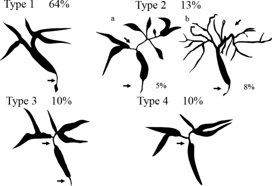
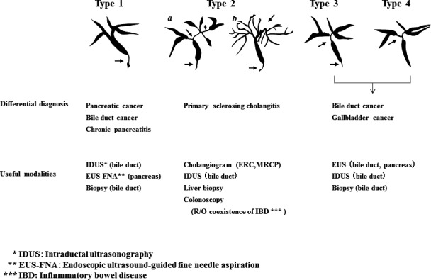
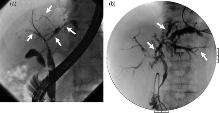
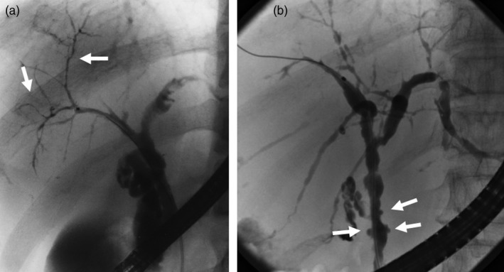
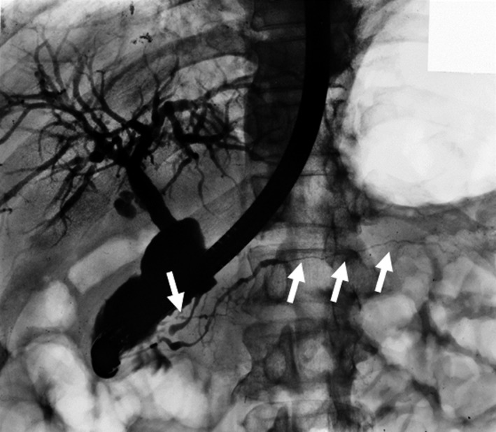
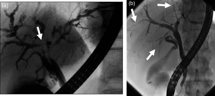
    1. Nakazawa T, Naitoh I, Hayashi K, Okumura F, Miyabe K, Yoshida M, et al. Diagnostic criteria for IgG4‐related sclerosing cholangitis based on cholangiographic classification. J Gastroenterol 2012;47:79–87. - PubMed
    1. Kamisawa T, Zen Y, Pillai S, Stone JH. IgG4‐related disease. Lancet. 2015;385:1460–71. - PubMed
    1. Ohara H, Okazaki K, Tsubouchi H, Inui K, Kawa S, Kamisawa T, et al. Clinical diagnostic criteria of IgG4‐related sclerosing cholangitis 2012. J Hepatobiliary Pancreat Sci. 2012;19:536–42. - PubMed
    1. Minds Guideline Center , Japan Council for Quality Health Care. Minds handbook for clinical practice guideline development 2014. 2014. Available from URL: https://minds.jcqhc.or.jp/s/guidance
    1. Fitch K, Bernstein SJ, Aguilar MS. The RAND/UCLA appropriateness method user's manual. Santa Monica: RAND; 2001.

Publication types

MeSH terms

Substances