. 2018 Dec 21;8(1):18024.
doi: 10.1038/s41598-018-36626-3.

Toll-Like Receptor-4 Disruption Suppresses Adipose Tissue Remodeling and Increases Survival in Cancer Cachexia Syndrome

Affiliations

Toll-Like Receptor-4 Disruption Suppresses Adipose Tissue Remodeling and Increases Survival in Cancer Cachexia Syndrome

Felipe Henriques et al. Sci Rep. .

Abstract

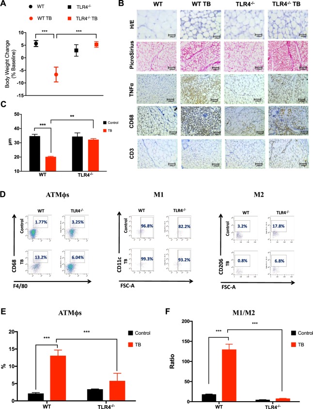
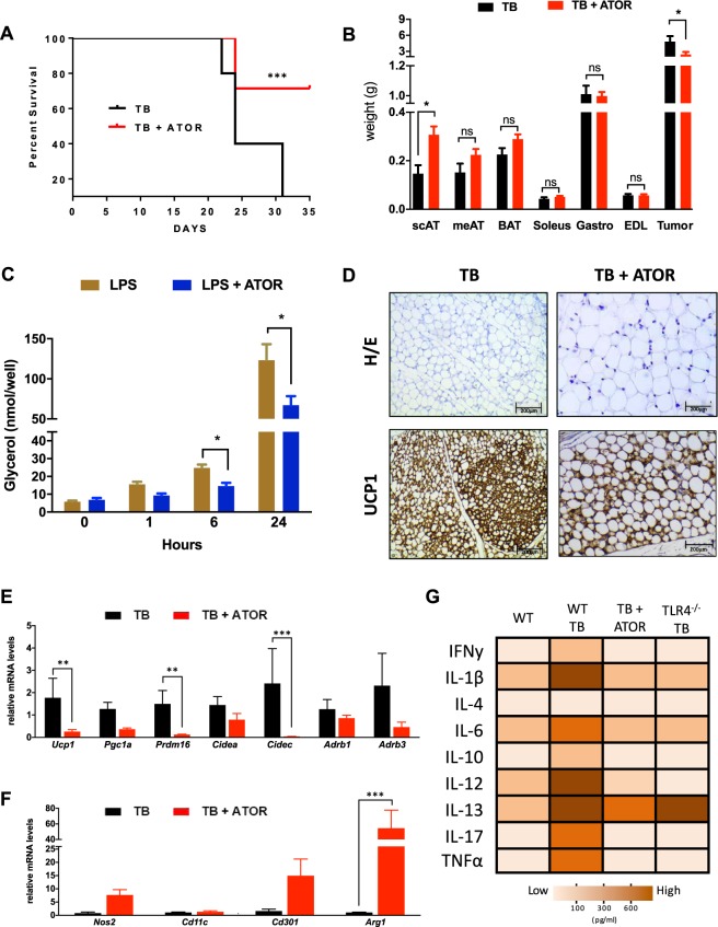
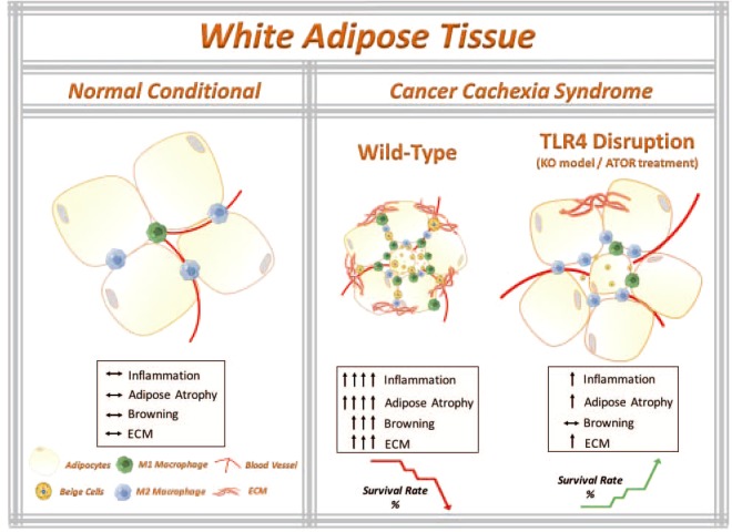
Cancer-induced cachexia, characterized by systemic inflammation, body weight loss, adipose tissue (AT) remodeling and muscle wasting, is a malignant metabolic syndrome with undefined etiology. Here, we show that both genetic ablation and pharmacological inhibition of TLR4 were able to attenuate the main clinical markers of cachexia in mice bearing Lewis lung carcinoma (LLC). AT remodelling was not found in LLC tumor-bearing (TB) TLR4-/- mice due to reduced macrophage infiltration and adipocyte atrophy. TLR4-/- mice were also resistant to cold-induced browning of subcutaneous AT (scAT). Importantly, pharmacological inhibition of TLR4 (Atorvastatin) reproduced the main protective effect against AT remodeling found in TLR4-/- TB mice. Moreover, the treatment was effective in prolonging survival and attenuating tumor mass growth when compared to non-treated-TB animals. Furthermore, tumor-induced elevation of circulating pro-inflammatory cytokines was similarly abolished in both genetic ablation and pharmacological inhibition of TLR4. These data suggest that TLR4 is a critical mediator and a promising target for novel anti-cachexia therapies.


Conflict of interest statement

The authors declare no competing interests.

Figures

Figure 1
Figure 1
TLR4 deletion attenuates scAT remodeling during cancer cachexia syndrome. (A) Wild-type C57BL/6 and TLR4−/− mice (8-week old male) were inoculated with LLC tumor cells. Body weight change (excluding tumor weight) was evaluated after 28 days before inoculated tumor cells. N = 10 per group. (B) Histologic sections of scAT in different experimental groups. Histological staining for H/E and picrosirius red were performed, and also, immunohistochemistry for inflammatory profile (TNFα) and immune cell markers (CD68 and CD3). N = 5 per group. (C) The size of adipocytes (cell diameter) from WT and TLR4−/− mice was quantitatively analyzed (500 adipocytes were measured for each group) after the experimental protocol. N = 5 per group. (D) Stromal vascular fractions (SVF) were isolated from scAT by collagenase digestion for each different group. Flow cytometric analysis of SVF was conducted using fluorescent-conjugated antibodies against CD68, F4/80, CD11c, CD206. Adipose tissue macrophages (ATMϕs) were defined as CD68+F4/80+ subpopulations and displayed the values as percentage of your respective groups. M1 and M2 ATMϕs were defined as CD68+F4/80+CD11c+CD206 and CD68+F4/80+CD11cCD206+, respectively. Representative flow cytometric dot plots showing the percentage of ATMϕs. N = 4 per group. (E) Quantification of double-positive cells for CD68+F4/80+ related to dot plots showed in (D). (F) M1/M2 ATMϕs ratio in the scAT. Scale bars, 100 μm and 200 μm. Graphs show the mean ± SEM. Statistical significance was determined by two-way ANOVA. **P < 0.01; ***P < 0.001.
Figure 2
Figure 2
Reduced triglyceride (TG) turnover is observed in TLR4−/− TB-mice. (A) Primary adipocytes from scAT were incubated in the presence (stimulated) or absence (basal) of isoproterenol (10−5M) for 1 hour. Graph shows lipolysis response assessed by the ratio of glycerol released (nmol/104 cells) relative to the basal condition. N = 4 per group. (B) Serum concentrations of non-esterified fatty acids (NEFA) and (C) % of palmitic acid in scAT were performed for each different group. N = 4 per group. (D) Immunoblot analysis for components of lipolysis. scAT lysates were immunoblotted for phospho-HSL (Ser660) and total HSL (E) Densitometric evaluation of phospho-HSL (Ser660) and total HSL. Ponceau staining was analyzed as a loading control. N = 4 per group. (F) qRT-PCR was performed to quantitate Pck1, Acsl1, Acads, Citrate Synthase, Me3 and Cpt1b mRNA levels in scAT from the different groups. N = 5 per group. Graph show the mean ± SEM. Statistical significance was determined by two-way ANOVA. *P < 0.01; ***P < 0.001.
Figure 3
Figure 3
TLR4 deletion reduces browning effect in TB mice throughout p38MAPK pathway. (A) Representative images of UCP1 staining of scAT from the different experimental groups. N = 5 per group. (B) Total quantification of UCP1 staining. (C) qRT-PCR was performed to quantitate Ucp1, Pgc1a, Prdm16, Cidea, Cidec, Adrb1 and Adrb3 mRNA levels in scAT from the different groups. N = 5 per group. (D) Depicted are representative immunoblots to detect phospho-p38MAPK, p38MAPK, phospho-AMPK and AMPK levels in scAT from the different experimental groups. N = 4 per group. (E) Densitometric evaluation of protein levels (phospho/total). Scale bars, 200 μm. Graphs show the mean ± SEM. Statistical significance was determined by two-way ANOVA. *P < 0.05; **P < 0.01; ***P < 0.001.
Figure 4
Figure 4
TLR4 is required for cold-induced browning phenotype. (A) Representative image of WT and TLR4−/− mice were single-caged and housed at room temperature (23 °C) and cold exposure (6 °C) for six days (N = 3 per genotype and per condition). (B) Representative image of UCP1 staining in scAT across the different genotypes. (C) Depicted are representative immunoblots to detect phospho-HSL, total HSL, UCP1, ATGL and Adiponectin. N = 3 per group. (D) Densitometric evaluation of phospho-HSL, total HSL, UCP1, ATGL and Adiponectin levels. (E) qRT-PCR was performed to quantitate Citrate synthase, Me3 and Cpt1b mRNA in scAT from the different groups. N = 3 per group. Scale Bars, 25 μm and 100 μm. Graph shows the mean ± SEM. Statistical significance was determined by one-way ANOVA. *P < 0.05; **P < 0.01. ***P < 0.001.
Figure 5
Figure 5
Atorvastatin treatment increases survival and improved cachexia-remodeling in scAT. (A) Kaplan-Meier survival curves show a statistically significant difference (P < 0.05) in survival between the Tumor Bearing (TB) mice (N = 15 per group) and Tumor Bearing + ATOR treatment (TB + ATOR) mice (N = 15 per group). (B) Adipose tissue, muscle and tumor weights at study end. (C) Time course of lipolysis in 3T3-L1 induced by LPS (100 ng) and LPS + ATOR treatment (100 μM). (D) Immunohistochemical analysis for UCP1 in scAT. (E) qRT-PCR was performed for Ucp1, Pgc1a, Prdm16, Cidea, Cidec, Adrb1 and  Adrb3 (F) Nos2, Cd11c, Cd301 and Arg1 (M1 and M2 macrophage polarization markers) for mRNA quantification in scAT from the different groups. N = 5 per group. (G) Heat map representing the circulating pro-inflammatory cytokines in the serum from the different experimental groups. White to brown scale depicts cytokine levels in pg/ml. Data were analyzed using the Bio-Plex manager software. N = 8 per group. Scale bars, 200 μm. Graph show the mean ± SEM. Significant differences were determined using Student’s t-test (B,E,F) and two-way ANOVA (C). Subcutaneous AT (scAT); Mesenteric AT (meAT); Brown AT (BAT); Extensor Digitorum Longus Muscle (EDL). *P < 0.05; **P < 0.01; ***P < 0.001.
Figure 6
Figure 6
The working model that TLR4 disruption ameliorates adipose tissue remodeling during cancer cachexia syndrome. Cancer-induced cachexia is characterized by systemic inflammation, body weight loss and AT remodeling. We show that TLR4 disruption (knockout model of TLR4 and pharmacological inhibition by ATOR treatment) ameliorates AT remodeling, in particular, preservation of adipocyte atrophy and attenuation of browning phenotype in scAT, as well as inflammatory responses during cancer cachexia syndrome. Additionally, TLR4 disruption was effective in prolonging the survival and reducing tumor mass growth. Therefore, these data suggest that TLR4 plays an important role during cachexia development. Here, we suggested a new potential therapeutic target for cancer cachexia syndrome. Parts of the figure were drawn using images from Servier Medical Art. Servier Medical Art by Servier is licensed under a Creative Commons Attribution 3.0 Unported License (https://smart.servier.com/).

References

    1. Fearon K, Arends J, Baracos V. Understanding the mechanisms and treatment options in cancer cachexia. Nat Rev Clin Oncol. 2013;10:90–99. doi: 10.1038/nrclinonc.2012.209. - DOI - PubMed
    1. Fearon K. Cachexia: Treat wasting illness on multiple fronts. Nature. 2016;529:156. doi: 10.1038/529156b. - DOI - PubMed
    1. Tsoli M, Swarbrick MM, Robertson GR. Lipolytic and thermogenic depletion of adipose tissue in cancer cachexia. Semin Cell Dev Biol. 2016;54:68–81. doi: 10.1016/j.semcdb.2015.10.039. - DOI - PubMed
    1. Batista ML, Jr., et al. Cachexia-associated adipose tissue morphological rearrangement in gastrointestinal cancer patients. J Cachexia Sarcopenia Muscle. 2016;7:37–47. doi: 10.1002/jcsm.12037. - DOI - PMC - PubMed
    1. Henriques FS, et al. Early suppression of adipocyte lipid turnover induces immunometabolic modulation in cancer cachexia syndrome. FASEB J. 2017;31:1976–1986. doi: 10.1096/fj.201601151R. - DOI - PubMed

Publication types

MeSH terms

LinkOut - more resources