. 2018 Dec 21;8(1):18021.
doi: 10.1038/s41598-018-36742-0.

Prickle1 regulates differentiation of frontal bone osteoblasts

Affiliations

Prickle1 regulates differentiation of frontal bone osteoblasts

Yong Wan et al. Sci Rep. .

Abstract

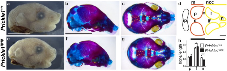
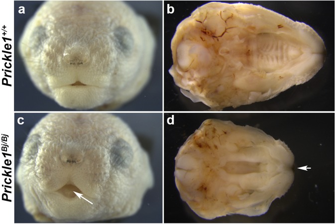
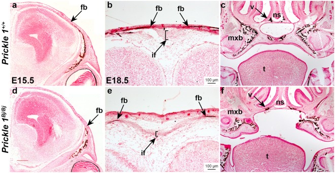
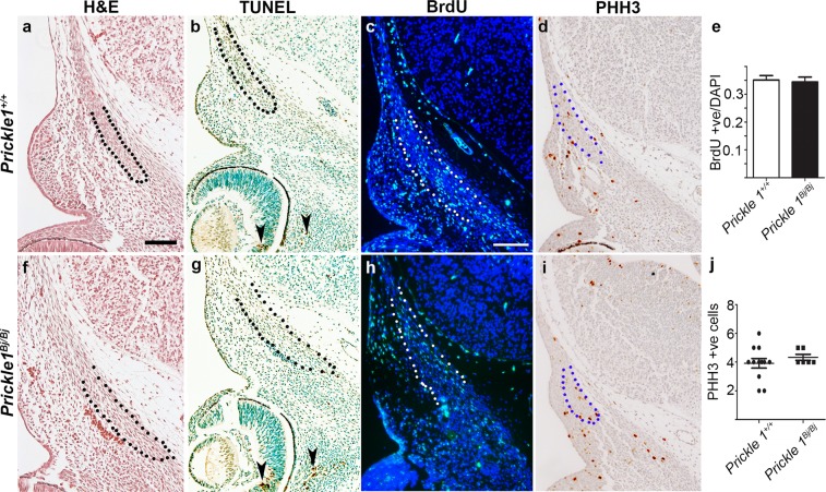
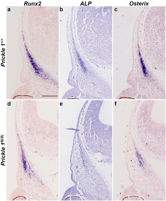
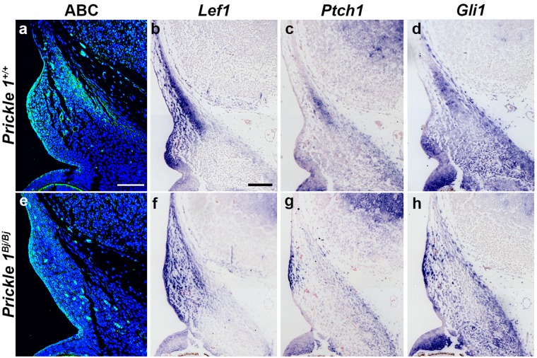
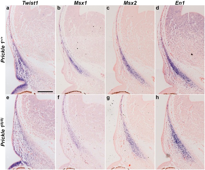
Enlarged fontanelles and smaller frontal bones result in a mechanically compromised skull. Both phenotypes could develop from defective migration and differentiation of osteoblasts in the skull bone primordia. The Wnt/Planar cell polarity (Wnt/PCP) signaling pathway regulates cell migration and movement in other tissues and led us to test the role of Prickle1, a core component of the Wnt/PCP pathway, in the skull. For these studies, we used the missense allele of Prickle1 named Prickle1Beetlejuice (Prickle1Bj). The Prickle1Bj/Bj mutants are microcephalic and develop enlarged fontanelles between insufficient frontal bones, while the parietal bones are normal. Prickle1Bj/Bj mutants have several other craniofacial defects including a midline cleft lip, incompletely penetrant cleft palate, and decreased proximal-distal growth of the head. We observed decreased Wnt/β-catenin and Hedgehog signaling in the frontal bone condensations of the Prickle1Bj/Bj mutants. Surprisingly, the smaller frontal bones do not result from defects in cell proliferation or death, but rather significantly delayed differentiation and decreased expression of migratory markers in the frontal bone osteoblast precursors. Our data suggests that Prickle1 protein function contributes to both the migration and differentiation of osteoblast precursors in the frontal bone.


Conflict of interest statement

The authors declare no competing interests.

Figures

Figure 1
Figure 1
Prickle1Bj/Bj mutants are microcephalic and have defects in the neural crest-derived skull. (ac,eg) Macroscopic views of Prickle1+/+ (ac) and Prickle1Bj/Bj (eg) littermates. (d) Schematic of the superior view of the skull vault. (h) quantification of skull vault measurements. (a,b,e,f) The Prickle1Bj/Bj mutant heads are shorter proximal-distally when observed laterally from the external (e) and alizarin red and alcian blue stained specimens (f). (c,g) Superior (birds-eye) view of the skull vault demonstrates that the interfrontal suture (yellow lines). (d) Schematic of the skull vault showing the tissue of origin and below is the schema for the measurements in (h). (h) The proximal-distal shortening of the Prickle1Bj/Bj mutants is most profound in the nasal region. c, coronal suture; f, frontal bone; if, interfrontal suture; ip, interparietal bone; m, mesoderm; n, nasal bone; ncc, neural-crest cell; p, parietal bone; s, sagittal suture. Scale bar = 0.5 cm.
Figure 2
Figure 2
Prickle1Bj/Bj mutants develop median cleft lip and cleft secondary palate. (a,c) Frontal views of wild-type (a) and Prickle1Bj/Bj (c) at P0. Median cleft lip indicated by arrow and is completely penetrant. (b,d) Palatal view of E18.5 fetuses. Arrow indicates medial cleft lip and cleft secondary palate.
Figure 3
Figure 3
Prickle1 protein is expressed in the craniofacial mesenchyme. We performed immunohistochemistry to Prickle1 protein at E12.5 (a,b) and E15.5 (df) and detected Prickle1 mRNA expression via Q-PCR at E15.5 (g). (a) Prickle1 protein is found in the frontal bone condensation at E12.5 (black outline). (b) A subset of the cells in the frontal bone express Prickle1 protein at E12.5 (black outline). (c) A horizontal section through the E15.5 skull showing von Kossa stained mineralized tissue in the frontal and parietal bones. (d) Section adjacent to c, stained with immunohistochemistry to Prickle1. (e) The E15.5 frontal bone expresses Prickle1 protein. (f) The E15.5 parietal bone also expresses Prickle1 protein. (e) At E15.5, Prickle1 mRNA expression is equivalent between the frontal and parietal bones (n = 7). Scale bars: a = 100 μm; b-f = 200 μm. cs, coronal suture; fb, frontal bone; pb, parietal bone.
Figure 4
Figure 4
Mineralization defects in the Prickle1Bj/Bj mutants. Histological analysis with von Kossa staining of Prickle1+/+ (ac) and Prickle1Bj/Bj (df) littermates at E15.5 (a,d) and E18.5 (b,c,e,f). (a,c) The smaller size of the Prickle1Bj/Bj frontal bones is apparent at E15.5 and the osteogenic fronts do not extend as far apically as the littermate control (black arrow). (b,e) The interfrontal suture is enlarged between the Prickle1Bj/Bj frontal bones (arrows). (c,f) The secondary palate is cleft and the maxillary and vomer (arrow) bones are displaced laterally. if, interfrontal suture; fb, frontal bone; mxb, maxillary bone; ns, nasal septum; t, tongue; v, vomer. Scale bar = 100 μm and applies to all.
Figure 5
Figure 5
No change in the rate of proliferation or apoptosis in the E12.5 Prickle1Bj/Bj frontal bones. E12.5 Prickle1+/+ (ad) and Prickle1Bj/Bj (fi) littermates assayed for histology (haemotoxylin and eosin) staining (a,f), TUNEL staining (b,g), proliferation with BrdU immunofluorescence (c,h) and mitosis with phospho-histone H3 immunohistochemistry (d,i). (a,d) The frontal bone mesenchymal condensation (black outline) is present in both wildtype (a) and Prickle1Bj/Bj (f) littermates. (b,g) TUNEL-positive cells were found near the eye (arrowheads) and absent in the frontal bone primordia. (c,h) BrdU-positive cells (green) are found in the frontal bone primordium (white outline) of wildtype (c) and Prickle1Bj/Bj (h) littermates. (d,i) Few positive PHH3-positive cells (brown) are found in the frontal bone primordium of wildtype (d) and Prickle1Bj/Bj (i) littermates. (e) No difference in the ratio of BrdU-positive cells in the frontal bone primordia (n = 3). (j) No difference in the number of PHH3-positive cells in the frontal bones between genotypes (n = 3). Scale bar = 100 μm.
Figure 6
Figure 6
Ossification is delayed in the frontal bone primordium. DIG-labeled section in situ hybridization to E12.5 Prickle1+/+ (ac) and Prickle1Bj/Bj (df) littermates. The expression levels of Runx2 (a,d), Alkaline phosphatase (ALP) (b,e) and Osterix (c,f) are decreased in frontal bone primordium of Prickle1Bj/Bj mutants compared with wild-type control embryos. Scale bar in a = 200 μm, and applies to all.
Figure 7
Figure 7
Wnt/β-catenin and Hedgehog signaling is decreased in the E12.5 frontal bone primordium. Immunofluorescence of active β-catenin (a,e) and DIG-labelled section in situ hybridization to Lef1 (b,f), Patched1 (Ptch1) (c, g) and Gli1 (d,h) in E12.5 Prickle1+/+ (ad) and Prickle1Bj/Bj (eh) littermates. (a,e) The level of active β-catenin (ABC) protein is decreased in the frontal bone primordium, but is maintained in the surface epithelium in the Prickle1Bj/Bj mutants compared with wild-type littermates. (b,f) Lef1 expression is reduced in the Prickle1Bj/Bj frontal bone primordium and throughout the mesenchyme surrounding it compared with wild-type littermates. (c,g) Ptch1 expression is reduced in the Prickle1Bj/Bj frontal bone primordium and throughout the mesenchyme surrounding it compared with control littermates. (d,h) Gli1 expression is also reduced in the Prickle1Bj/Bj frontal bone primordium compared with control littermates. Scale bar = 100 μm.
Figure 8
Figure 8
Osteoblast migration is decreased in the frontal bone primordium. DIG-labeled section in situ hybridization to Twist1, Msx1, Msx2 and Engrailed1 (En1) to E12.5 Prickle1+/+ (ad) and Prickle1Bj/Bj (eh) coronal sections. (a,e) Decreased expression of Twist1 in the Prickle1Bj/Bj compared with wild-type. (b,f) The expression of Msx1 is decreased in the mutant compared with wild-type. (c,g) The expression of Msx2 is slightly decreased in the Prickle1Bj/Bj compared with wild-type. (d,h) The expression of En1 is similar in the Prickle1Bj/Bj compared with wild type. Scale bar in a = 200 μm, and applies to all.

References

    1. McBratney-Owen B, Iseki S, Bamforth SD, Olsen BR, Morriss-Kay GM. Development and tissue origins of the mammalian cranial base. Developmental biology. 2008;322:121–132. doi: 10.1016/j.ydbio.2008.07.016. - DOI - PMC - PubMed
    1. Jiang X, Iseki S, Maxson RE, Sucov HM, Morriss-Kay GM. Tissue origins and interactions in the mammalian skull vault. Developmental biology. 2002;241:106–116. doi: 10.1006/dbio.2001.0487. - DOI - PubMed
    1. Yoshida T, Vivatbutsiri P, Morriss-Kay G, Saga Y, Iseki S. Cell lineage in mammalian craniofacial mesenchyme. Mechanisms of development. 2008;125:797–808. doi: 10.1016/j.mod.2008.06.007. - DOI - PubMed
    1. Merrill AE, et al. Cell mixing at a neural crest-mesoderm boundary and deficient ephrin-Eph signaling in the pathogenesis of craniosynostosis. Human molecular genetics. 2006;15:1319–1328. doi: 10.1093/hmg/ddl052. - DOI - PubMed
    1. Bialek P, et al. A twist code determines the onset of osteoblast differentiation. Developmental cell. 2004;6:423–435. doi: 10.1016/S1534-5807(04)00058-9. - DOI - PubMed

Publication types

MeSH terms

Substances

LinkOut - more resources