. 2019 Apr;38(16):3003-3018.
doi: 10.1038/s41388-018-0638-9. Epub 2018 Dec 21.

Loss of ER retention motif of AGR2 can impact mTORC signaling and promote cancer metastasis

Affiliations

Loss of ER retention motif of AGR2 can impact mTORC signaling and promote cancer metastasis

Katrin Tiemann et al. Oncogene. 2019 Apr.

Abstract

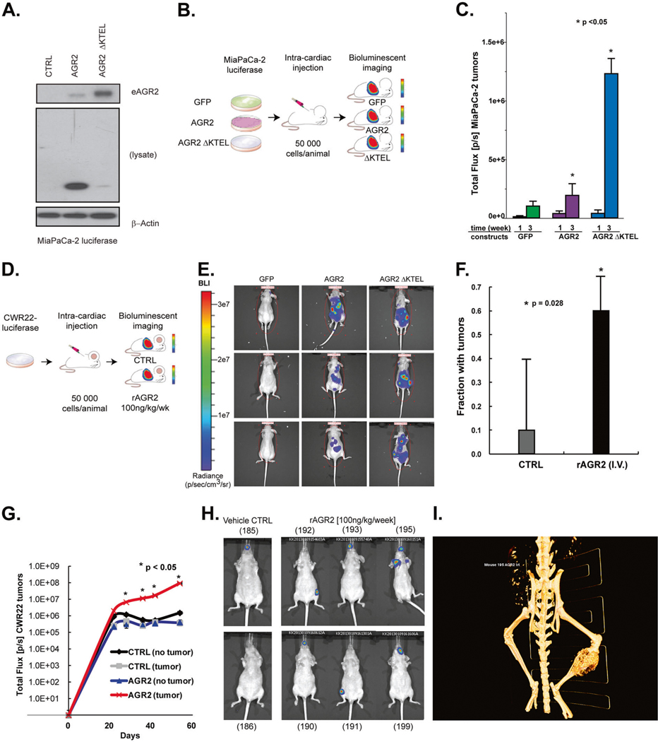
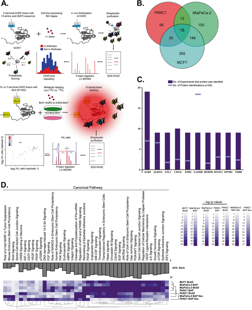
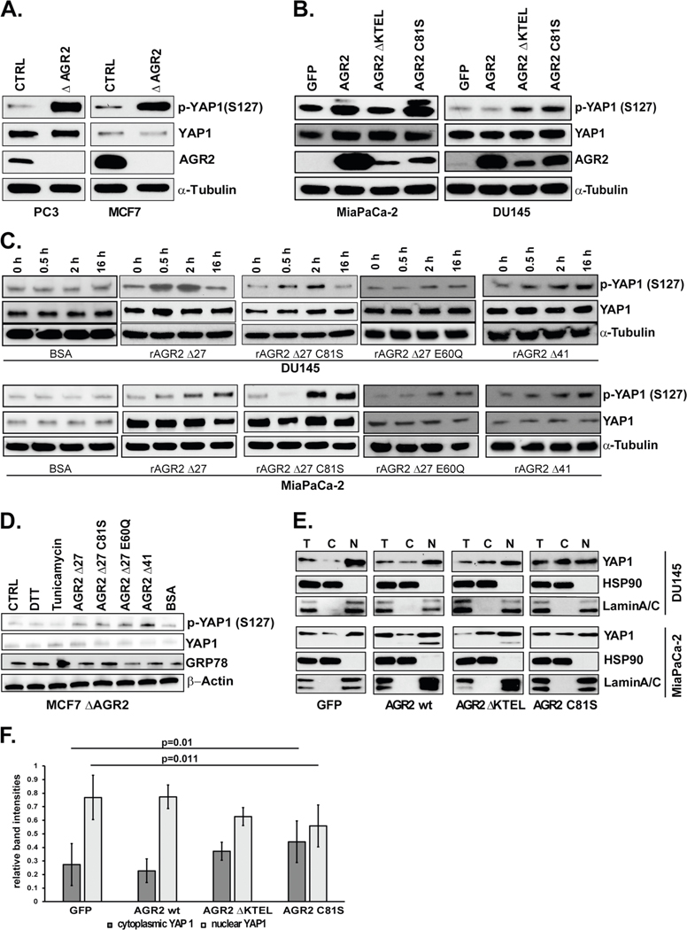
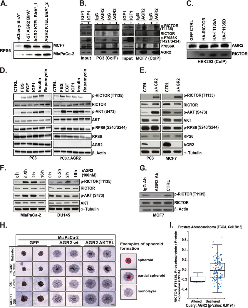
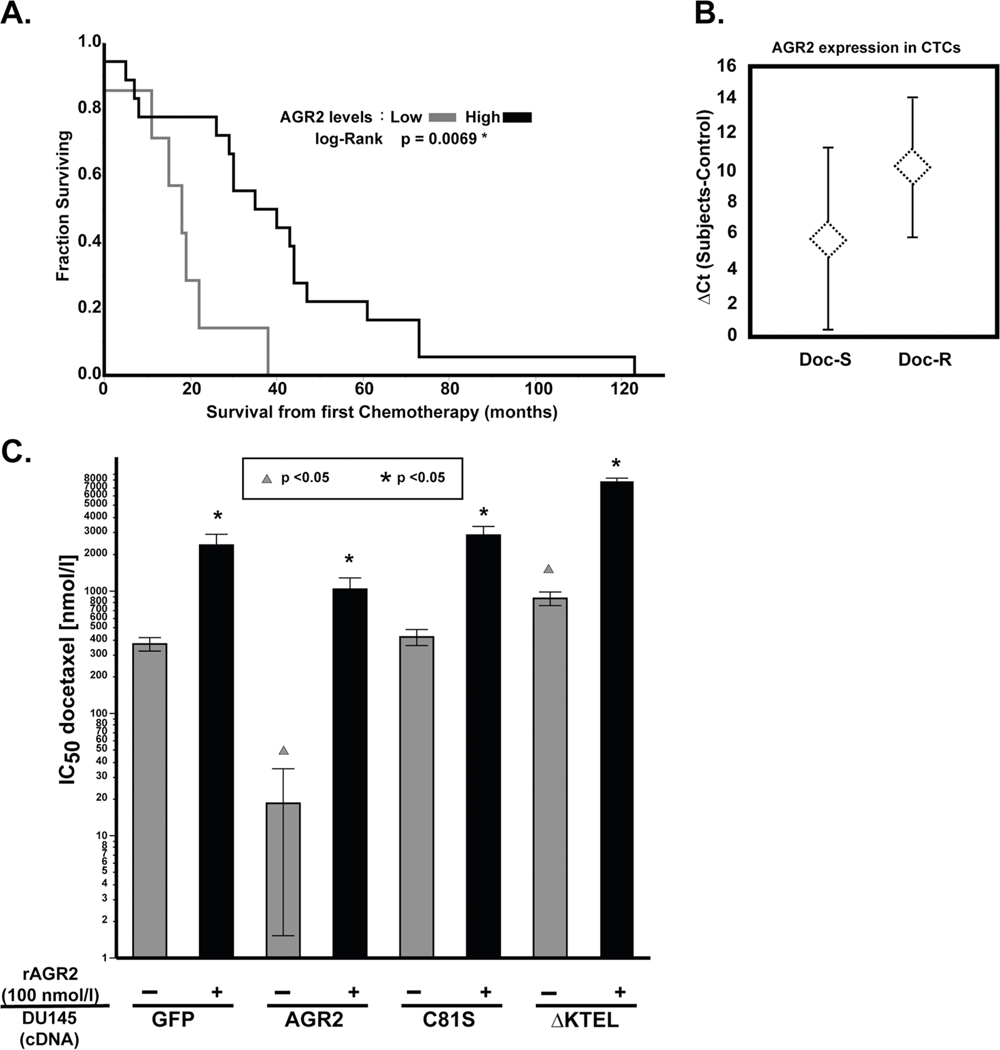
Anterior gradient 2 (AGR2) is a member of the protein disulfide isomerase (PDI) family, which plays a role in the regulation of protein homeostasis and the unfolded protein response pathway (UPR). AGR2 has also been characterized as a proto-oncogene and a potential cancer biomarker. Cellular localization of AGR2 is emerging as a key component for understanding the role of AGR2 as a proto-oncogene. Here, we provide evidence that extracellular AGR2 (eAGR2) promotes tumor metastasis in various in vivo models. To further characterize the role of the intracellular-resident versus extracellular protein, we performed a comprehensive protein-protein interaction screen. Based on these results, we identify AGR2 as an interacting partner of the mTORC2 pathway. Importantly, our data indicates that eAGR2 promotes increased phosphorylation of RICTOR (T1135), while intracellular AGR2 (iAGR2) antagonizes its levels and phosphorylation. Localization of AGR2 also has opposing effects on the Hippo pathway, spheroid formation, and response to chemotherapy in vitro. Collectively, our results identify disparate phenotypes predicated on AGR2 localization. Our findings also provide credence for screening of eAGR2 to guide therapeutic decisions.


Conflict of interest statement

Conflict of interest The authors declare that they have no conflict of interest.

Figures

Fig. 1
Fig. 1
eAGR2 promotes cancer metastasis in prostate and pancreatic cancer xenografts. A Loss of ER retention sequence (KTEL) increases levels of eAGR2 in spent media in MiaPaCa-2 cells. B MiaPaCa-2 pancreatic cancer cells carrying luciferase were transfected with GFP, AGR2 wt, and AGR2 ΔKTEL and were injected into left ventricle of (N = 10) nude mice (per arm) to investigate the impact of increased secretion of AGR2 and tumor growth. C Error bars are expressed as average ± SD. Results were normally distributed with equal variance between groups were analyzed by unpaired two-tailed Student’s t test. Asterisks denote p value significance (Student’s t test, p < 0.05) between AGR2 wt and AGR2 ΔKTEL. D Prostate cancer xenografts (N = 10 per arm) were injected with purified recombinant AGR2 by intravenous injection in the left ventricle show increased tumor incidence and E, F with higher flux G, H than control treated mice. Error bars are expressed as average ± SD. Results were normally distributed with equal variance between two groups and were analyzed by paired two-tailed Student’s t test. Asterisks denote p value significance (p < 0.05). i Metastatic spread to right leg (red arrow) but not the left leg (gray arrow) was observed in two mice by CT scan (mouse #195)
Fig. 2
Fig. 2
Interrogation of AGR2-interacting partners in the ER and extracellular space. A We performed immunoprecipitation assays followed by mass spectrometry (MS) analysis of BAP-AGR2 and AGR2-BAP. Protein lists were submitted to CRAPome (contaminant repository for affinity purification) and SAINT (significance analysis of INTeractome) [54] analysis, which allowed the identification of high-scoring interactions. We also used proximity ligation of biotin to prey proteins by the expression of bait proteins fused to a nonspecific biotin ligase, BirA* (BioID). In vivo biotinylation of proximal proteins was detected by streptavidin-based affinity purification and proteomics identification. Isotopic labeling of cells with control (BirA* mCherry) transfected cells was used to refine proteins in the AGR2 interactome. B Total number of AGR2-interacting proteins after removal of contaminants. C AGR2 was identified in (n = 19/19) experiments derived from whole-cell lysates (although its expression levels were lower than high abundant contaminant proteins) (GAPDH). D New biological pathways ascribed to AGR2 were enriched by ingenuity pathway analysis. The AGR2 interactome was enriched for proteins that have been linked to the mTORC pathway in all three cancer cell lines used in this assay. We also noted that analysis of the AGR2 interactome in the extracellular (Secreted) space clustered differently that the whole-cell lysates (whole-cell lysates)
Fig. 3
Fig. 3
eAGR2 alters activation of Hippo pathway via phosphorylation of YAP1. A Knockout of AGR2 in PC3 and MCF7 cell lines. Lysates were analyzed by immunoblot. B Overexpression of GFP, AGR2 wt, AGR2 ΔKTEL, and AGR2 C81S in MiaPaCa-2 and DU145 cells Lysates were analyzed by immunoblot. C Treatment of DU145 and MiaPaCa-2 cell lines with 100 nmol/l rAGR2 Δ27, rAGR2 Δ27 C81S, rAGR2 Δ27 E60Q, and rAGR2 Δ41 over 0.5, 2, and 16 h. Totally, 100 nmol/l of BSA was used as control. D MCF7 ΔAGR2 cells were treated with 1 mM DTT, 100 nmol/l BSA, 100 nmol/l rAGR2 or 5 μmol/l Tunicamycin for 2 h. Untreated cells were used as control (CTRL). Expression of UPR marker GRP78, p-YAP1, YAP1, and β-Actin were determined by western blot. E Nuclear and cytosolic fractionation of DU145 and MiaPaCa-2 lysates transfected with GFP and wt AGR2, AGR2 Δ KTEL, or AGR2 C81S were analyzed by immunoblot. F Quantitation of the immunoblots of five independent fractionation experiments (ImageJ) of DU145 cells. A two-tailed Student’s t test was used to assess statistical significance (p = 0.01 and p = 0.011). All immunoblots are representative of data from at least three independent biological replicates
Fig. 4
Fig. 4
AGR2 interacts with mTORC2 complex thereby altering its signaling and cellular phenotype. A Stably transfected MCF7 and MiaPaCa-2 cells with AGR2 BioID constructs were subject to streptavidin pulldown and subsequently analyzed by immunoblot for RPS6 interaction with AGR2. As control mCherry BirA* was used. B Co-Immunoprecipitation (CoIP) of endogenous AGR2 or isotype matched IgG in PC3 and MCF7 cells under serum starvation (CTRL) or treated with 50 nmole/l IGF1 for 2 h. Cell lysates corresponding to input and CoIP fractions were subject to immunoblot for RICTOR/p-RICTOR, p-P79S6K/P70S6K with AGR2. C CoIP of transient transfected HEK293 cells with HA-RICTOR, HA-RICTOR-1135A, and HARICTOR-1135D cDNA. Cell lysates were incubated with biotinylated AGR2 (6 h) and precipitated with streptavidin magnetic beads. Immunoblot of the elute was probed for AGR2 and RICTOR. D PC3 and PC3 ΔAGR2 cell lines were serum starved or treated with 10% FBS, EGF (25 ng/ml), IGF1 (40 ng/ml), Insulin (250 nmol/l), and Rapamycin (20 nmol/l) for 2 h. The lysates were subjected to immunoblot for RICTOR, AKT, and RPS6. E Whole-cell lysates corresponding to knockout of AGR2 in PC3 and MCF7 cells were subject to immunoblot for mTORC2 (RICTOR) and its downstream components. F Time-course of MiaPaCa-2 and DU145 cells treated with rAGR2 and probed for RICTOR and AKT. G Treatment of MCF7 cells with AGR2 neutralizing antibody (500 nmole/l) or IgG (500 nmole/l) for 24 h. Lysates were subject to immunoblot of RICTOR. H Spheroid formation assay of pancreatic cancer cell lines transfected cells with GFP, AGR2 wt, and AGR2 DKTEL while treated with (PBS/untreated), rAGR2 with and without OSI 1 μmole/l for 48 h postplating. Spheriod formation was imaged by microscopy. An example of spheroid initiation formation is indicated by the drawn circles around the denser spheroid area. Area outside are cells not participating yet. I Correlation of AGR2 expression and phosphorylation of RICTOR in independent cohorts of patients with pancreatic (TCGA) and prostate adenocarcinomas [44] (unpaired Student’s t test). All in vitro experiments represent at least three independent biological replicates
Fig. 5
Fig. 5
Loss of ER retention motif promotes contralateral tumor growth. A Prostate cancer xenografts were used to assess the impact of juxtacrine AGR2 on contralateral tumor growth (n = 8). B DU145 mouse tumors at site 1 contralateral to AGR2 ΔKTEL are significantly larger (p < 0.05) than wildtype tumors contralateral to AGR2 or AGR2 C81S (Student’s t test, n = 8). Site 2 tumors show no significant differences in size. C Two dissected tumors of site 1 (GFP tumor) collateral toAGR2 ΔKTEL, AGR2 C81S, and AGR2 wt. The tumor location, mouse number, and weight of tumor are displayed
Fig. 6
Fig. 6
Clinical significance of AGR2 expression in prostate cancer patients treated with chemotherapy. A Prostate cancer patients with high-AGR2 mRNA levels (median) have significantly longer survival from first chemotherapy (Mann–Whitney–Wilcoxon test, p = 0.0069). B AGR2 expression is increased in CTC-enriched peripheral blood (7.5 ml) obtained from CRPC patients prior to initiating docetaxel treatment the mean AGR2 expression (±SD) from CTC-enriched RNA in docetaxel-sensitive (Doc-S, n = 15) and docetaxel-resistant (Doc-R, n = 7) subjects were examined by RT-PCR. Docetaxel response is defined as a ≥30% reduction in PSA at 12 weeks as suggested by PSAWG2 guidelines [55]. We observe a strong trend towards increased AGR2 expression in the Doc-R cohort, which approaches statistical significance (Student’s t test, p = 0.06). C DU145 parental or DU145 transfected with GFP, ΔKTEL, or C81S were treated with docetaxel for 48 h and treated with rAGR2 (100 nmol/l). Cell viability was assessed by MTS assay and IC50 was determined. Comparison of dosing with rAGR2 (100 nmol/l) is statistically represented by asterisks (Student’s t test, p < 0.05). Comparison of transfection with AGR2 cDNA constructs (GFP as the reference) is statistically represented by triangles (Student’s t test, p < 0.05). All experiments represent at least three independent biological replicates

References

    1. Liu D, Rudland PS, Sibson DR, Platt-Higgins A, Barraclough R.Human homologue of cement gland protein, a novel metastasis inducer associated with breast carcinomas. Cancer Res. 2005;65:3796–805. - PubMed
    1. Patel P, Clarke C, Barraclough DL, Jowitt TA, Rudland PS, Barraclough R, et al. Metastasis-promoting anterior gradient 2 protein has a dimeric thioredoxin fold structure and a role in cell adhesion. J Mol Biol. 2012;425:929–43. - PubMed
    1. Park SW, Zhen G, Verhaeghe C, Nakagami Y, Nguyenvu LT, Barczak AJ, et al. The protein disulfide isomerase AGR2 is essential for production of intestinal mucus. Proc Natl Acad Sci USA. 2009;106:6950–5. - PMC - PubMed
    1. Higa A, Mulot A, Delom F, Bouchecareilh M, Nguyen DT, Boismenu D, et al. Role of pro-oncogenic protein disulfide isomerase (PDI) family member anterior gradient 2 (AGR2) in the control of endoplasmic reticulum homeostasis. J Biol Chem. 2011;286:44855–68. - PMC - PubMed
    1. Clarke C, Rudland P, Barraclough R. The metastasis-inducing protein AGR2 is O-glycosylated upon secretion from mammary epithelial cells. Mol Cell Biochem. 2015;408:245–52. - PMC - PubMed

Publication types

MeSH terms