. 2019 Mar 1;76(3):301-309.
doi: 10.1001/jamaneurol.2018.4053.

Clinical, Radiologic, and Prognostic Features of Myelitis Associated With Myelin Oligodendrocyte Glycoprotein Autoantibody

Affiliations

Clinical, Radiologic, and Prognostic Features of Myelitis Associated With Myelin Oligodendrocyte Glycoprotein Autoantibody

Divyanshu Dubey et al. JAMA Neurol. .

Abstract

Importance: Recognizing the characteristics of myelin oligodendrocyte glycoprotein autoantibody (MOG-IgG) myelitis is essential for early accurate diagnosis and treatment.

Objective: To evaluate the clinical, radiologic, and prognostic features of MOG-IgG myelitis and compare with myelitis with aquaporin-4-IgG (AQP4-IgG) and multiple sclerosis (MS).

Design, setting, and participants: We retrospectively identified 199 MOG-IgG-positive Mayo Clinic patients from January 1, 2000, through December 31, 2017, through our neuroimmunology laboratory. Fifty-four patients met inclusion criteria of (1) clinical myelitis; (2) MOG-IgG positivity; and (3) medical records available. We excluded 145 patients without documented myelitis. Myelitis of AQP4-IgG (n = 46) and MS (n = 26) were used for comparison.

Main outcomes and measures: Outcome variables included modified Rankin score and need for gait aid. A neuroradiologist analyzed spine magnetic resonance imaging of patients with MOG-IgG and control patients blinded to diagnosis.

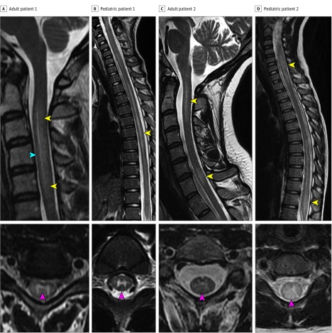
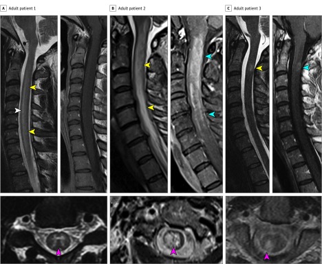
Results: Of 54 included patients with MOG-IgG myelitis, the median age was 25 years (range, 3-73 years) and 24 were women (44%). Isolated transverse myelitis was the initial manifestation in 29 patients (54%), and 10 (19%) were initially diagnosed as having viral/postviral acute flaccid myelitis. Cerebrospinal fluid-elevated oligoclonal bands occurred in 1 of 38 (3%). At final follow-up (median, 24 months; range, 2-120 months), 32 patients (59%) had developed 1 or more relapses of optic neuritis (n = 31); transverse myelitis (n = 7); or acute disseminated encephalomyelitis (n = 1). Clinical features favoring MOG-IgG myelitis vs AQP4-IgG or MS myelitis included prodromal symptoms and concurrent acute disseminated encephalomyelitis. Magnetic resonance imaging features favoring MOG-IgG over AQP4-IgG or MS myelitis were T2-signal abnormality confined to gray matter (sagittal line and axial H sign) and lack of enhancement. Longitudinally extensive T2 lesions were of similar frequency in MOG-IgG and AQP4-IgG myelitis (37 of 47 [79%] vs 28 of 34 [82%]; P = .52) but not found in MS. Multiple spinal cord lesions and conus involvement were more frequent with MOG-IgG than AQP4-IgG but not different from MS. Wheelchair dependence at myelitis nadir occurred in one-third of patients with MOG-IgG and AQP4-IgG but never with MS, although patients with MOG-IgG myelitis recovered better than those with AQP4-IgG.

Conclusions and relevance: Myelitis is an early manifestation of MOG-IgG-related disease and may have a clinical phenotype of acute flaccid myelitis. We identified a variety of clinical and magnetic resonance imaging features that may help clinicians identify those at risk in whom MOG-IgG should be tested.


Conflict of interest statement

Conflict of Interest Disclosures: Dr Pittock and Mayo Clinic have a financial interest in patents #12/678350 (filed 2010) and #12/573942 (filed 2008) that relate to functional aquaporin-4 (AQP4)/neuromyelitis optica (NMO)–IgG assays and NMO-IgG as a cancer marker; Dr Pittock has provided consultation to Alexion Pharmaceuticals, Medimmune, and Chugai Pharma USA but has received no personal fees or personal compensation for these consulting activities. All compensation for consulting activities is paid directly to Mayo Clinic. Dr Pittock has received a research grant from Alexion Pharmaceuticals for an investigator-initiated study as well as support from the National Institutes of Health (RO1 NS065829-01) and the Guthy Jackson Charitable Foundation for NMO research. Dr Weinshenker receives royalties from RSR Ltd, Oxford University, Hospices Civil de Lyon, and MVZ Labor PD Volkmann und Kollegen for a patent of NMO-IgG as a diagnostic test for NMO and related disorders; serves as a member of an adjudication committee for clinical trials in NMO being conducted by MedImmune and Alexion pharmaceutical companies; is a consultant for Caladrius Biosciences and Brainstorm Therapeutics regarding potential clinical trials for NMO; and serves as a member of a data safety monitoring committee for clinical trials conducted by Novartis. Dr Lucchinetti receives grant support from Novartis, Sanofi-Synthelabo, Biogen, Mallinkrodt, and Alexion. Dr McKeon has received research support from MedImmune and Euroimmun. Dr Keegan has had research funded by Biogen and receives publishing royalties for Common Pitfalls in Multiple Sclerosis and CNS Demyelinating Diseases. Dr Tillema receives grant support from the National Institutes of Health (NCATS; KL2 TR000136). Dr Patterson has research funded by Orphazyme; has received grant support from National Institutes of Health (U54NS065768); receives royalties from Elsevier (Encyclopedia of the Neurological Sciences), Up-To-Date (Section Editor, Pediatric Neurology), and Sage Publishing (Journal of Child Neurology and Child Neurology Open); receives a stipend as editor-in-chief of the Journal of Child Neurology and Child Neurology Open and travel expenses as editor of the Journal of Inherited Metabolic Disease and Journal of Inherited Metabolic Disease Reports; has served as a member of data safety monitoring committees for clinical trials conducted by Novartis and Amicus; and has served as a consultant for Actelion, Alexion, Orphazyme, Shire, and Vtesse. Dr Messacar receives grant support from the National Institutes of Health (1K23AI128069-01). Dr Tyler receives grant support from the National Institutes of Health (R01NS101208). Dr Flanagan is a site principal investigator in a randomized placebo-controlled clinical trial of inebilizumab in neuromyelitis optica spectrum disorders funded by MedImmune/Viela Bio.

Figures

Figure 1.
Figure 1.. Examples of Magnetic Resonance Imaging (MRI) Findings in Myelin Oligodendrocyte Glycoprotein Autoantibody (MOG-IgG) Myelitis
A, An adult patient with a sagittal T2-hyperintense line (blue arrowhead) that is surrounded by fainter T2 hyperintensity (yellow arrowheads), with a corresponding T2 hyperintensity highly restricted to the gray matter on axial sequences forming an H sign (red arrowhead). B, A pediatric patient with a sagittal line of T2 hyperintensity (yellow arrowhead) confined to gray matter on axial images forming an H sign (red arrowhead). C, An adult patient with 2 noncontiguous T2-hyperintense lesions in the cervical cord extending 2.5 and 1 vertebral segments (yellow arrowhead); the upper lesion has a vague appearance of a central line. The axial sequences show central T2 hyperintensity most prominent at the central canal and fainter signal surrounding that is confined to gray matter vaguely resembling an H sign (red arrowhead). D, A pediatric patient is shown with 2 longitudinally extensive T2 hyperintense lesions on sagittal sequences, 1 in the cervical cord and 1 in the conus (yellow arrowheads). On axial sequences, this lesion shows central predominant signal abnormality that is more diffuse and not confined to gray matter (red arrowhead).
Figure 2.
Figure 2.. Comparison of Myelitis With Myelin Oligodendrocyte Glycoprotein Autoantibody (MOG-IgG), Aquaporin-4–IgG (AQP4-IgG), and Multiple Sclerosis (MS) Myelitis
A, An adult patient with MOG-IgG myelitis is shown in which a sagittal T2-hyperintense line (white arrowhead) is present surrounded by hazier T2 hyperintensity (yellow arrowheads) forming a longitudinally extensive spinal cord lesion accompanied by mild spinal cord swelling. On axial sequences, the T2 hyperintensity is highly restricted to the gray matter forming an H sign (red arrowhead). Postgadolinium sagittal images demonstrate no gadolinium enhancement. B, In contrast, an adult patient with AQP4-IgG positive neuromyelitis optica spectrum disorder (NMOSD) and a longitudinally T2 hyperintense extensive lesion with prominent swelling (yellow arrowheads) and an axial T2 lesion shows diffuse central signal abnormality not confined to gray matter (red arrowhead). The lesion has accompanying gadolinium enhancement (blue arrowheads). C, An adult patient with MS has a short T2-hyperintense lesion at the C2 level extending one vertebral segment in length (yellow arrowhead) with accompanying faint ring enhancement (blue arrowhead). On axial sequences, the lesion is located in the periphery of the cord in the right dorsal column (red arrowhead).

References

    1. Greenberg BM. Treatment of acute transverse myelitis and its early complications. Continuum (Minneap Minn). 2011;17(4):733-743. - PubMed
    1. Zalewski NL, Flanagan EP, Keegan BM. Evaluation of idiopathic transverse myelitis revealing specific myelopathy diagnoses. Neurology. 2018;90(2):e96-e102. - PubMed
    1. Jiao Y, Fryer JP, Lennon VA, et al. . Updated estimate of AQP4-IgG serostatus and disability outcome in neuromyelitis optica. Neurology. 2013;81(14):1197-1204. doi:10.1212/WNL.0b013e3182a6cb5c - DOI - PMC - PubMed
    1. Lennon VA, Kryzer TJ, Pittock SJ, Verkman AS, Hinson SR. IgG marker of optic-spinal multiple sclerosis binds to the aquaporin-4 water channel. J Exp Med. 2005;202(4):473-477. doi:10.1084/jem.20050304 - DOI - PMC - PubMed
    1. Reindl M, Di Pauli F, Rostásy K, Berger T. The spectrum of MOG autoantibody-associated demyelinating diseases. Nat Rev Neurol. 2013;9(8):455-461. doi:10.1038/nrneurol.2013.118 - DOI - PubMed

MeSH terms