

Origin, Organization, Dynamics, and Function of Actin and Actomyosin Networks at the T Cell Immunological Synapse
- PMID: 30576253
- PMCID: PMC8343269
- DOI: 10.1146/annurev-immunol-042718-041341
Origin, Organization, Dynamics, and Function of Actin and Actomyosin Networks at the T Cell Immunological Synapse
Abstract
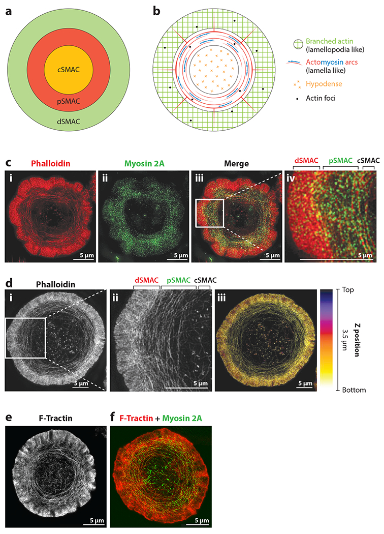
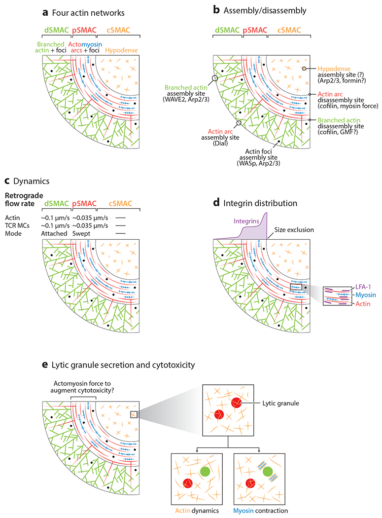
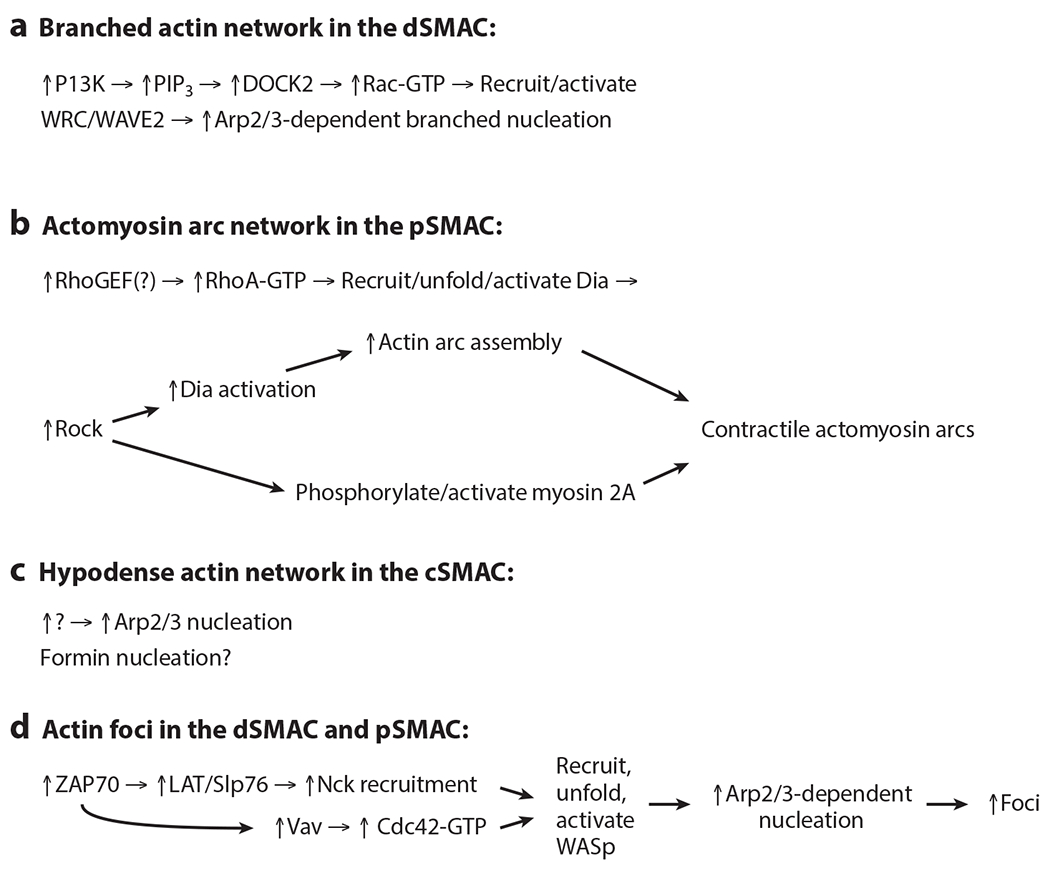
The engagement of a T cell with an antigen-presenting cell (APC) or activating surface results in the formation within the T cell of several distinct actin and actomyosin networks. These networks reside largely within a narrow zone immediately under the T cell's plasma membrane at its site of contact with the APC or activating surface, i.e., at the immunological synapse. Here we review the origin, organization, dynamics, and function of these synapse-associated actin and actomyosin networks. Importantly, recent insights into the nature of these actin-based cytoskeletal structures were made possible in several cases by advances in light microscopy.
Keywords: T cell; actin; formin; immunological synapse; lytic granule; myosin.
Figures
Similar articles
-
Formin-generated actomyosin arcs propel T cell receptor microcluster movement at the immune synapse.J Cell Biol. 2016 Nov 7;215(3):383-399. doi: 10.1083/jcb.201603080. Epub 2016 Oct 31. J Cell Biol. 2016. PMID: 27799367 Free PMC article.
-
Actin retrograde flow and actomyosin II arc contraction drive receptor cluster dynamics at the immunological synapse in Jurkat T cells.Mol Biol Cell. 2012 Mar;23(5):834-52. doi: 10.1091/mbc.E11-08-0731. Epub 2012 Jan 4. Mol Biol Cell. 2012. PMID: 22219382 Free PMC article.
-
Cytoskeletal tension actively sustains the migratory T-cell synaptic contact.EMBO J. 2020 Mar 2;39(5):e102783. doi: 10.15252/embj.2019102783. Epub 2020 Jan 2. EMBO J. 2020. PMID: 31894880 Free PMC article.
-
Exploring How Cytoskeleton Dynamics Tunes T Cell Activation at the Immunological Synapse.Eur J Immunol. 2025 Aug;55(8):e70028. doi: 10.1002/eji.70028. Eur J Immunol. 2025. PMID: 40770849 Review.
-
The role of actomyosin and the microtubular network in both the immunological synapse and T cell activation.Front Biosci. 2007 Jan 1;12:437-47. doi: 10.2741/2073. Front Biosci. 2007. PMID: 17127308 Review.
Cited by
-
Comparative Transcriptome Sequencing Analysis of Hirudo nipponia in Different Growth Periods.Front Physiol. 2022 Jun 23;13:873831. doi: 10.3389/fphys.2022.873831. eCollection 2022. Front Physiol. 2022. PMID: 35812329 Free PMC article.
-
LFA-1 and kindlin-3 enable the collaborative transport of SLP-76 microclusters by myosin and dynein motors.J Cell Sci. 2021 Aug 15;134(16):jcs258602. doi: 10.1242/jcs.258602. Epub 2021 Aug 27. J Cell Sci. 2021. PMID: 34279667 Free PMC article.
-
A B-cell actomyosin arc network couples integrin co-stimulation to mechanical force-dependent immune synapse formation.Elife. 2022 Apr 11;11:e72805. doi: 10.7554/eLife.72805. Elife. 2022. PMID: 35404237 Free PMC article.
-
A novel method for characterizing cell-cell interactions at single-cell resolution reveals unique signatures in blood T cell-monocyte complexes during infection.bioRxiv [Preprint]. 2024 Sep 23:2024.09.20.612103. doi: 10.1101/2024.09.20.612103. bioRxiv. 2024. PMID: 39386643 Free PMC article. Preprint.
-
Mechanical regulation of lymphocyte activation and function.J Cell Sci. 2024 Jul 1;137(13):jcs219030. doi: 10.1242/jcs.219030. Epub 2024 Jul 12. J Cell Sci. 2024. PMID: 38995113 Free PMC article. Review.
References
-
- Cannon JL, Burkhardt JK. 2002. The regulation of actin remodeling during T-cell-APC conjugate formation. Immunol. Rev 186(1):90–99 - PubMed
-
- Huang Y, Burkhardt JK. 2007. T-cell-receptor-dependent actin regulatory mechanisms. J. Cell Sci 120(5):723–30 - PubMed
-
- Billadeau DD, Nolz JC, Gomez TS. 2007. Regulation of T-cell activation by the cytoskeleton. Nat. Rev. Immunol 7(2):131–43 - PubMed
-
- Burkhardt JK, Carrizosa E, Shaffer MH. 2008. The actin cytoskeleton in T cell activation. Annu. Rev. Immunol 26(1):233–59 - PubMed
Publication types
MeSH terms
Substances
Grants and funding
LinkOut - more resources
Full Text Sources
