. 2018 Dec 21;13(12):e0208791.
doi: 10.1371/journal.pone.0208791. eCollection 2018.

Physiological relevance of epithelial geometry: New insights into the standing gradient model and the role of LI cadherin

Affiliations

Physiological relevance of epithelial geometry: New insights into the standing gradient model and the role of LI cadherin

Yana Vereshchaga et al. PLoS One. .

Abstract

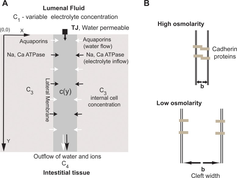
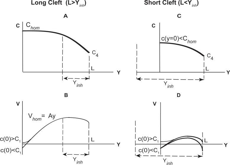
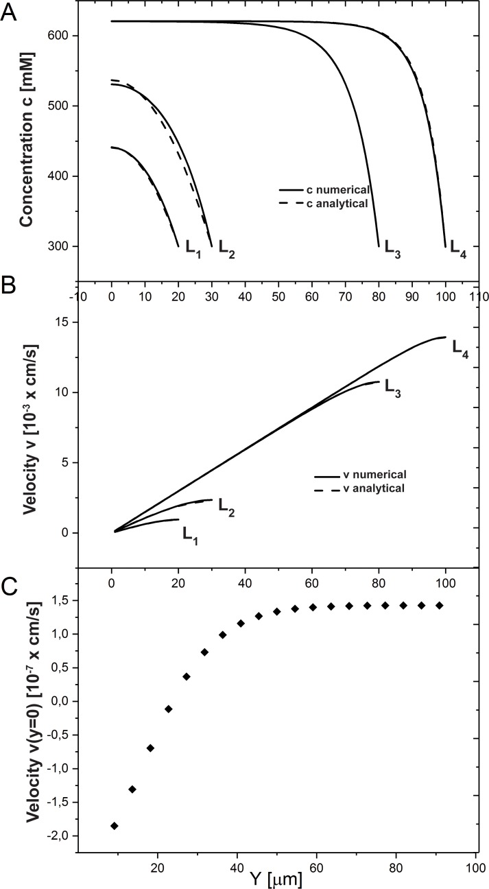
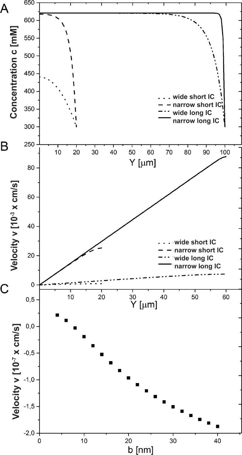
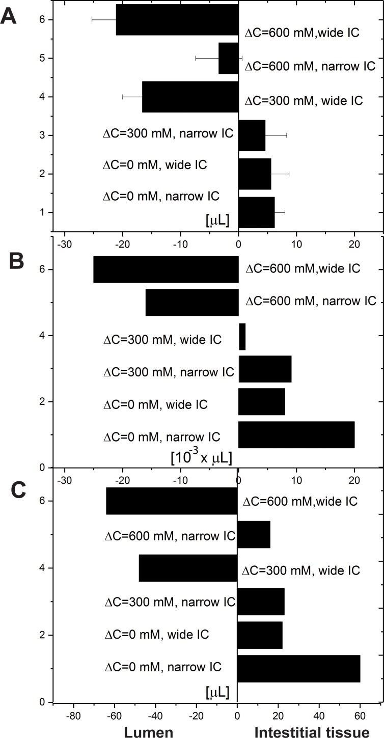
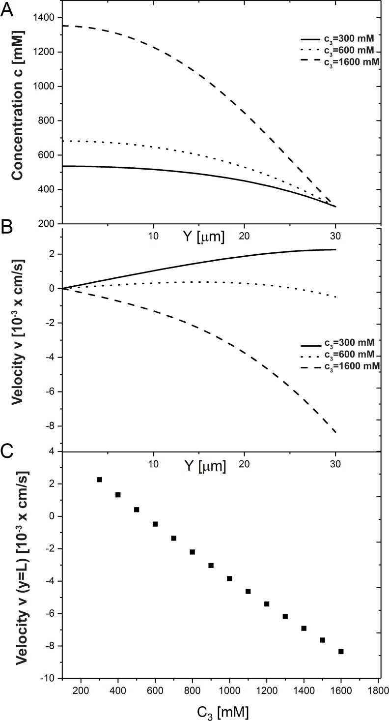
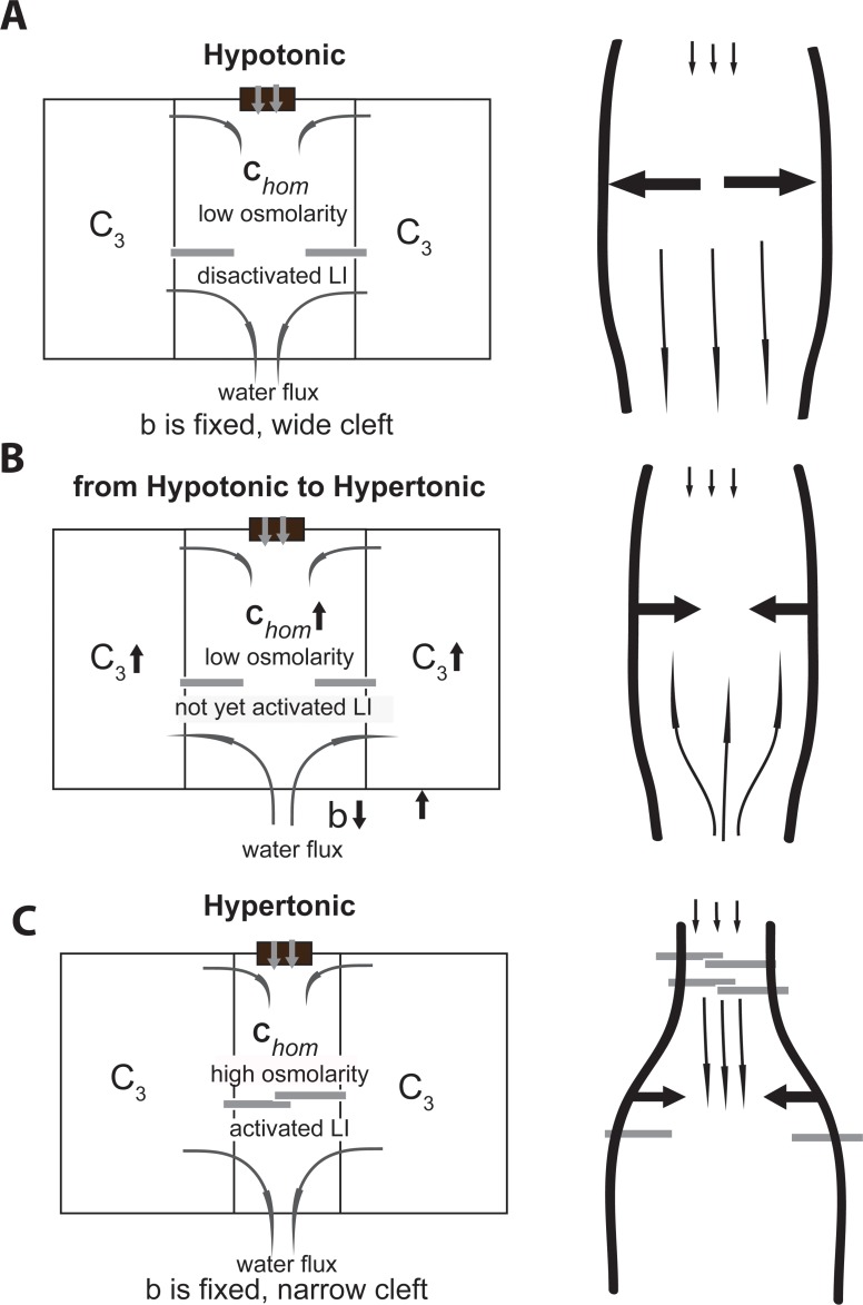
We introduce a mathematical model of an absorbing leaky epithelium to reconsider the problem formulated by Diamond and Bossert in 1967: whether "… some distinctive physiological properties of epithelia might arise as geometrical consequences of epithelial ultrastructure". A standing gradient model of the intercellular cleft (IC) is presented that includes tight junctions (TJ) and ion channels uniformly distributed along the whole cleft. This nonlinear system has an intrinsic homogeneous concentration and the spatial scale necessary to establish it along the cleft. These parameters have not been elucidated so far. We further provide non-perturbative analytical approximations for a broad range of parameters. We found that narrowing of the IC increases ion concentration dramatically and can therefore prevent outflow through tight junctions (TJs) and the lateral membrane, as long as extremely high luminal osmolarities are not reached. Our model predicts that the system is to some extent self-regulating and thereby prevents fluxes into the lumen. Recent experimental evidence has shown that liver-intestine (LI) cadherin can control the up/down flux in intestines via regulation of the cleft width. This finding is in full agreement with predictions of our model. We suggest that LI-cadherin may increase water transport through epithelia via sequential narrowing of the cleft, starting from the highest concentration area at the beginning of the cleft and triggering a propagating squeezing motion.


Conflict of interest statement

The authors have declared that no competing interests exist.

Figures

Fig 1
Fig 1. Model of water and electrolyte transport through simple epithelia and IC width regulation via LI-cadherins.
a) The model comprises four compartments, which are (1) the lumen of the organ (e.g. the gut), (2) the lateral intercellular cleft (IC), where the values of c(y), v(y), p(y) are calculated, (3) the cytoplasm of the cell, and (4) the interstitial tissue. In the lumen a given concentration of electrolytes is assumed. The tight junctions (TJ) separate the lumen (1) and the IC (2), and are assumed to be impermeable to the electrolyte and permeable to water with a permeability coefficient kTJ. The concentration of electrolytes in the cytoplasm is assumed to be constant c3 (except in the case in which we explored its influence on the direction of water flow). ATPases are assumed to pump the electrolyte through the lateral membrane into the lateral intercellular cleft. The interstitial tissue is assumed to exhibit a constant electrolyte concentration c4, which is maintained by the blood vessels located there. The important compartment is the IC. Water enters this compartment through the TJ, aquaporins or from the interstitial tissue. Ions enter through the lateral membrane due to the ATPases and leave the IC due to diffusion and due to the water flux, flushing the lateral intercellular cleft. b) The width of the lateral intercellular cleft b depends on the binding activity of the 7D-cadherins, which in turn depends on the extracellular Ca2+ level. High Ca2+ concentration triggers LI-cadherin binding and low one courses protein disruption. In the publication [12] we reported the results with a designed peptide that disrupts the LI-cadherins in the epithelium CACO2 and the cleft becomes broader as a result.
Fig 2
Fig 2
Schematical graphs of the ion concentration c and water velocity v in long (L>yinh) (A-B) and short (Lyinh) (C-D) clefts. Physiologically related scenarious are shown. Non-physiological ones, for example, when c(L) is much higher than spatially homogeneous concentration chom, are not shown here.
Fig 3
Fig 3. Influence of cleft length on ion concentration (c) and water velocity (v).
Concentration (A) and velocity (B) profiles (analytical approximation and numerical solutions (see S1 File for details)) in the case of long (L>yinh,L3 = 80 μm,L4 = 100 μm) and short (Lyinh, L1 = 20 μm, L2 = 30 μm) clefts are shown. (C) Change in velocity at the cleft TJ end. Increase in cleft length reverses the water flux in the TJ from negative to positive. The following system parameters were used: C3 = 290 mM, C4 = 300 mM, C1 = 600 mM.
Fig 4
Fig 4. Influence of cleft width on ion concentration and water velocity.
(A) The concentration increases as the cleft width gets decreased from 400 nm (wide IC) to 40 nm (narrow IC). The results for a short L = 20 μmyinh and long L = 100 μm>yinh cleft are shown. (B) The velocity growth is shown for short and long clefts (L = 60 μm>yinh) as IC width decreases. (C) The flux direction (velocity v(0)) through the TJ alters from negative to positive one as IC becomes wider. The following system parameters were used: C3 = 290 mM, C4 = 300 mM.
Fig 5
Fig 5. Change in water volume per hour for strong, medium hypertonic, and isotonic osmolarities in the lumen.
Osmolarity difference between lumen and interstitial (Δc = c1c4) is accordingly 600, 300, and 0 mM. The results are presented for wide (b = 400 nm) and narrow (b = 40 nm) clefts. (A) Experimentally measured fluxes, as the peptide disrupts LI cadherins binding (the IC widening as shown in Fig 1B with low osmolarity). The figure is adapted from [12]; (B) Water flux change through the TJ, numerical results; (C) Water flux through the open end of the IC as a result of elevated cytosolic concentration c3. Changes in response to the alteration of lumen osmolarity: 900mM, 600mM and 300mM. The following system parameters were used: c4 = 300 mM,L = 20 μm.
Fig 6
Fig 6. Influence of internal cell concentration c3.
(A) Ion concentration and (B) water velocity profiles along the IC. (C) The velocity at the end of the cleft v(L) as c3 increases from 200 to 1600 mM. The IC length is L = 30mM, and c4 = 300mM.
Fig 7
Fig 7. The potential mechanism by which LI-cadherins can squeeze water out of the IC in the course of lumen osmolarity change is introduced schematically.
(A) The cleft is wide as LI-cadherins are in deactivated state. (B) Change from hypotomic to hypertonic conditions. LI-cadherins are not yet activated, the internal cell c3 and the homogeneous concentrations increase. Water flux in the negative direction and through lateral membrane is present. (C) LI-cadherins are activated as the homogeneous concentration chom reaches a certain value–LI activation causes the water to be squeezed downwards.

Similar articles

Cited by

References

    1. Reid EW. Transport of fluid by certain epithelia. The Journal of Physiology. 1901; 26: 436–444. 10.1113/jphysiol.1901.sp000844 - DOI - PMC - PubMed
    1. Curran PF, Solomon AK. Ion and water fluxes in the ileum of rats. J Gen Physiol. 1957; 594 41: 143–168. - PMC - PubMed
    1. Windhager EE, Whittembury G, Oken DE, Schatzmann HJ, Solomon AK. Single proximal 596 tubules of the Necturus kidney. III. Dependence of H2O movement on NaCl concentration. 597 Am J Physiol. 1959; 197: 313–318. 10.1152/ajplegacy.1959.197.2.313 - DOI - PubMed
    1. Diamond JM. The mechanism of isotonic water transport. J Gen Physiol. 1964; 48: 15–42. - PMC - PubMed
    1. Parsons DS, Wingate DL. The effect of osmotic gradients on fluid transfer across rat 600 intestine in vitro. Biochim Biophys Acta. 1961; 46: 170–183. - PubMed

Publication types