Review
. 2018 Dec 20;8(1):3.
doi: 10.3390/jcm8010003.

Innate Immune Responses and Viral-Induced Neurologic Disease

Affiliations
Review

Innate Immune Responses and Viral-Induced Neurologic Disease

Yuting Cheng et al. J Clin Med. .

Abstract

Multiple sclerosis (MS) is a disease of the central nervous system (CNS) characterized by chronic neuroinflammation, axonal damage, and demyelination. Cellular components of the adaptive immune response are viewed as important in initiating formation of demyelinating lesions in MS patients. This notion is supported by preclinical animal models, genome-wide association studies (GWAS), as well as approved disease modifying therapies (DMTs) that suppress clinical relapse and are designed to impede infiltration of activated lymphocytes into the CNS. Nonetheless, emerging evidence demonstrates that the innate immune response e.g., neutrophils can amplify white matter damage through a variety of different mechanisms. Indeed, using a model of coronavirus-induced neurologic disease, we have demonstrated that sustained neutrophil infiltration into the CNS of infected animals correlates with increased demyelination. This brief review highlights recent evidence arguing that targeting the innate immune response may offer new therapeutic avenues for treatment of demyelinating disease including MS.

Keywords: demyelination; innate immunity; neutrophils; remyelination; virus.


Conflict of interest statement

The authors declare no conflicts of interest.

Figures

Figure 1
Figure 1
Derivation and characterization of a mouse model in which CXCL1 expression within the central nervous system (CNS) is under the control of a doxycycline promoter. (A) Cartoon depiction of experimental strategy to generate double (dbl) transgenic (tg) mice in which expression of mouse CXCL1 is under control of the glial fibrillary acidic protein (GFAP) promoter upon doxycycline treatment. (B) Cortex tissue from double tg and single tg postnatal day 1 (P1) mice was dissociated and enriched for astrocytes. Following 24-h of Dox (100 ng/mL) treated double tg astrocyte cultures, immunofluorescence confirmed CXCL1 expression within GFAP-positive astrocytes while vehicle treatment yielded no CXCL1 fluorescence (original magnification, ×20). (C) Within the spinal cord (SC) and brain, dox-treated double tg mice had statistically significant increases in CXCL1 mRNA expression over Dox-treated single tg mice at days 7 and 12 post-infection (p.i.) * p < 0.05, ** p < 0.01. Data derived from Marro et al., (2016) [101].
Figure 2
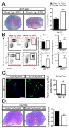
Figure 2
Elevated CNS CXCL1 expression is associated with increased neutrophil accumulation and demyelination. (A) Representative luxol fast blue (LFB)-stained spinal cords reveals increased (p < 0.05) demyelination in mouse hepatitis virus (JHMV)-infected Dox-treated double tg mice compared to single tg controls. (B) Flow cytometric analysis revealed a significant increase in the frequency and total number of neutrophils within the spinal cord of JHMV-infected Dox-treated double tg mice compared to single tg mice. (C) Representative immune-fluorescence staining further demonstrated a significant increase in the number of Ly6B.2-positive neutrophils (yellow arrowheads) within the spinal cord parenchyma of JHMV-infected double tg compared to single tg mice; red arrowheads indicate neutrophils located within the spinal cord meninges. Quantification of neutrophils within the spinal cords indicated an overall increase (p < 0.05) in Dox-treated double tg mice compared to Dox-treated single tg mice. (D) Representative LFB-stained spinal cord sections from JHMV-infected double tg mice treated with either control IgG2a or anti-Ly6G antibody between days 3 to 15 p.i. Quantification of the severity of demyelination revealed reduced white matter damage in mice treated with anti-Ly6G antibody compared to mice treated with isogenic IgG2a control antibody. * p < 0.05, ** p < 0.01, *** p < 0.001. Data derived from Marro et al., (2016) [101].
Figure 3
Figure 3
Characterization of possible neutrophil mechanisms of action that contribute to white matter damage. Cartoon depiction of neutrophil mechanisms of action including release of reactive nitrogen species (RNS) [105] and reactive oxygen species (ROS) [106] intermediates, neutrophil extracellular traps (NETs) [83], and select cytokines [107]. We hypothesize that these mechanisms may be responsible for the enhanced white matter damage observed following induced infiltration of neutrophils in preclinical mouse models of MS.

References

    1. Steinman L. Immunology of Relapse and Remission in Multiple Sclerosis. Annu. Rev. Immunol. 2014;32:257–281. doi: 10.1146/annurev-immunol-032713-120227. - DOI - PubMed
    1. Lassmann H., Bruck W., Lucchinetti C.F. The immunopathology of multiple sclerosis: An overview. Brain Pathol. 2007;17:210–218. doi: 10.1111/j.1750-3639.2007.00064.x. - DOI - PMC - PubMed
    1. Neumann H., Medana I.M., Bauer J., Lassmann H. Cytotoxic T lymphocytes in autoimmune and degenerative CNS diseases. Trends Neurosci. 2002;25:313–319. doi: 10.1016/S0166-2236(02)02154-9. - DOI - PubMed
    1. Steinman L. Multiple sclerosis: A coordinated immunological attack against myelin in the central nervous system. Cell. 1996;85:299–302. doi: 10.1016/S0092-8674(00)81107-1. - DOI - PubMed
    1. Greenfield A.L., Hauser S.L. B-cell Therapy for Multiple Sclerosis: Entering an era. Ann. Neurol. 2018;83:13–26. doi: 10.1002/ana.25119. - DOI - PMC - PubMed