Review
. 2018 Dec 20;11(1):5.
doi: 10.3390/cancers11010005.

Glioblastoma: Microenvironment and Niche Concept

Affiliations
Review

Glioblastoma: Microenvironment and Niche Concept

Davide Schiffer et al. Cancers (Basel). .

Abstract

The niche concept was originally developed to describe the location of normal neural stem cells (NSCs) in the subependymal layer of the sub-ventricular zone. In this paper, its significance has been extended to the location of tumor stem cells in glioblastoma (GB) to discuss the relationship between GB stem cells (GSCs) and endothelial cells (ECs). Their interaction is basically conceived as responsible for tumor growth, invasion and recurrence. Niches are described as the points of utmost expression of the tumor microenvironment (TME), therefore including everything in the tumor except for tumor cells: NSCs, reactive astrocytes, ECs, glioma-associated microglia/macrophages (GAMs), myeloid cells, pericytes, fibroblasts, etc. and all intrinsic and extrinsic signaling pathways. Perivascular (PVNs), perinecrotic (PNNs) and invasive niches were described from the pathological point of view, highlighting the basic significance of the EC/tumor stem cell couple. PNN development was reinterpreted based on the concept that hyperproliferative areas of GB are composed of GSCs/progenitors. TME was depicted in its function as the main regulator of everything that happens in the tumor. A particular emphasis was given to GAMs, pericytes and reactive astrocytes as important elements affecting proliferation, growth, invasion and resistance to therapies of tumor cells.

Keywords: glioblastoma; microenvironment; niche; pericytes; reactive astrocytes.


Conflict of interest statement

The authors declare no conflict of interest.

Figures

Figure 1
Figure 1
Glioblastoma, IDH wild type. (a) CD34+ endothelial cells of arterioles and capillaries in direct contact with Nestin+ tumor cells; double immunostaining with CD34 (DAB) and Nestin (Fast Red), original magnification (OM) ×400. (b) Vessel co-option. Sleeve of tumor cells around capillaries; CD34, DAB, OM ×400. (c) Id., with several Nestin+ tumor cells; DAB, OM ×200. (d) Mild infiltration. Reactive astrocytes on small vessels; GFAP, DAB, OM ×200; (e) More intense infiltration. Reactive astrocytes on vessels; GFAP, DAB, OM ×200; (f) High infiltration. Reactive astrocytes with end-feet on small vessels; GFAP, DAB, OM ×400. IDH, isocitrate dehydrogenase; DAB, 3,3′-Diaminobenzidine.
Figure 2
Figure 2
Glioblastoma, IDH wild type. (a) Mild infiltration with inital vessel increase; H&E, original magnification (OM) ×200. (b) Id., CD34+ endothelial cells; DAB, OM ×200. (c) More advanced tumor infiltration, small vessel with endothelial proliferation and sprouts; H&E, OM ×400. (d) Id., CD34+ endothelial cells; DAB, OM ×400. (e) Mild infiltration with perivascular macrophages; CD163, DAB, OM ×200. (f) Infiltration area with leaked perivascular macrophages; CD163, DAB, OM ×200. (g) Ramified microglia in tumor parenchyma and perivascular macrophages; Iba-1, DAB, OM ×200. (h) Proliferated tumor vessels with NG2/CSPG4+ endothelial cells and pericytes; DAB, OM ×200. (i) Glomeruli with α-SMA pericytes; DAB, OM ×200. IDH, isocitrate dehydrogenase; H&E, hematoxylin and eosin; DAB, DAB, 3,3′-Diaminobenzidine.
Figure 3
Figure 3
Glioblastoma, IDH wild type. (a) Circumscribed necrosis in a hyperproliferative area; H&E, original magnification (OM) ×200. The hyperproliferative zone bordering necrosis is almost GFAP-negative; DAB, OM ×200 (b) and ×400 (c). The same area is highly Nestin-positive; DAB, OM ×200 (d) and ×400 (e). The same area shows a high Ki-67/MIB-1 labeling index; DAB, OM ×200 (f) and ×400 (g). The same area is highly Sox2-positive; DAB, OM ×200 (h) and ×400 (i). The same area is positive for Musashi-1, cryostat section, immunofluorescence (green) (j) and highly CD133-positive, cryostat sections, immunofluorescence (red), both OM ×400 (k). IDH, isocitrate dehydrogenase; H&E, hematoxylin and eosin; DAB, DAB, 3,3′-Diaminobenzidine [49].
Figure 4
Figure 4
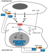
Relationship between a stem cell/progenitor and an endothelial cell [13].
Figure 5
Figure 5
(a) Development of circumscribed necrosis. (b) Equilibrium between tumor non-stem cells and tumor stem cells [66].

Similar articles

Cited by

References

    1. Stupp R., Mason W.P., van den Bent M.J., Weller M., Fisher B., Taphoorn M.J., Belanger K., Brandes A.A., Marosi C., Bogdahn U., et al. Radiotherapy plus concomitant and adjuvant temozolomide for glioblastoma. N. Engl. J. Med. 2005;352:987–996. doi: 10.1056/NEJMoa043330. - DOI - PubMed
    1. Holland E.C. Progenitor cells and glioma formation. Curr. Opin. Neurol. 2001;14:683–688. doi: 10.1097/00019052-200112000-00002. - DOI - PubMed
    1. Schiffer D., Mellai M., Annovazzi L., Casalone C., Cassoni P. Tumor Microenvironment: Perivascular and Perinecrotic Niches in Gliomas. In: Morgan L.R., editor. Molecular Considerations and Evolving Surgical Management Issues in the Treatment of Patients with a Brain Tumor. 1st ed. InTech; Rjieka, Croatia: 2015. pp. 49–82.
    1. Sanai N., Alvarez-Buylla A., Berger M.S. Neural stem cells and the origin of gliomas. N. Engl. J. Med. 2005;353:811–822. doi: 10.1056/NEJMra043666. - DOI - PubMed
    1. Assanah M., Lochhead R., Ogden A., Bruce J., Goldman J.J., Canoll P. Glial progenitors in adult white matter are driven to form malignant gliomas by platelet-derived growth factor-expressing retroviruses. J. Neurosci. 2006;26:6781–6790. doi: 10.1523/JNEUROSCI.0514-06.2006. - DOI - PMC - PubMed

LinkOut - more resources