. 2018 Dec 22;18(1):199.
doi: 10.1186/s12862-018-1309-8.

Classification, substrate specificity and structural features of D-2-hydroxyacid dehydrogenases: 2HADH knowledgebase

Affiliations

Classification, substrate specificity and structural features of D-2-hydroxyacid dehydrogenases: 2HADH knowledgebase

Dorota Matelska et al. BMC Evol Biol. .

Erratum in

Abstract

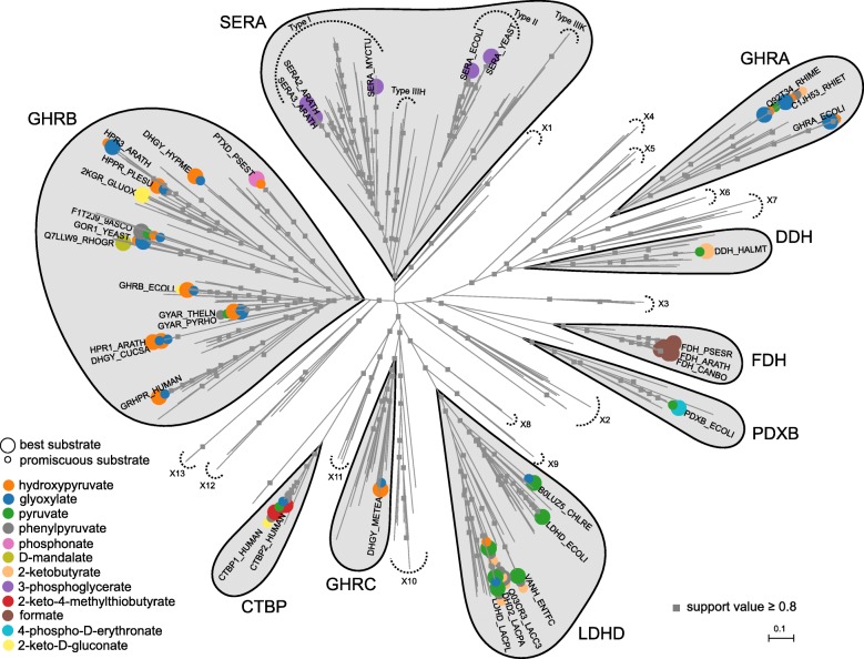
Background: The family of D-isomer specific 2-hydroxyacid dehydrogenases (2HADHs) contains a wide range of oxidoreductases with various metabolic roles as well as biotechnological applications. Despite a vast amount of biochemical and structural data for various representatives of the family, the long and complex evolution and broad sequence diversity hinder functional annotations for uncharacterized members.

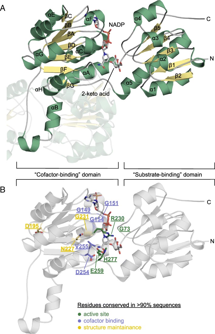
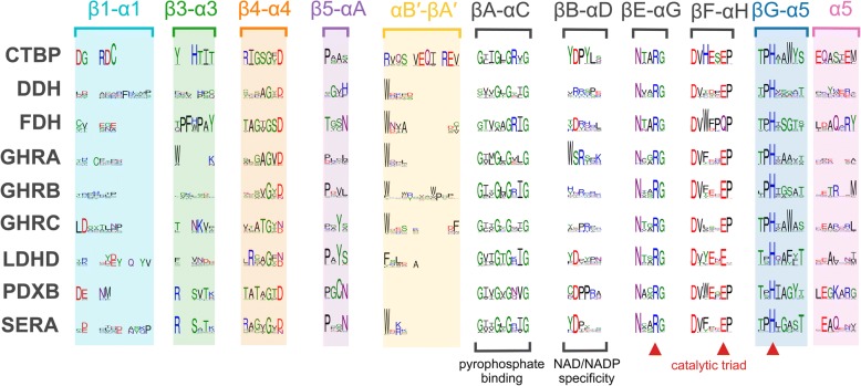
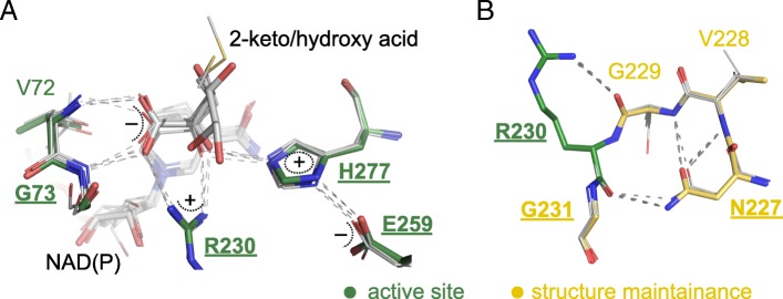
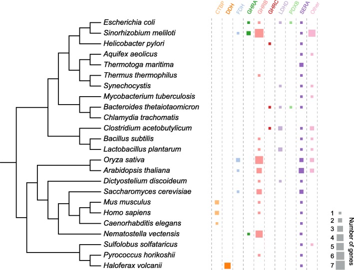
Results: We report an in-depth phylogenetic analysis, followed by mapping of available biochemical and structural data on the reconstructed phylogenetic tree. The analysis suggests that some subfamilies comprising enzymes with similar yet broad substrate specificity profiles diverged early in the evolution of 2HADHs. Based on the phylogenetic tree, we present a revised classification of the family that comprises 22 subfamilies, including 13 new subfamilies not studied biochemically. We summarize characteristics of the nine biochemically studied subfamilies by aggregating all available sequence, biochemical, and structural data, providing comprehensive descriptions of the active site, cofactor-binding residues, and potential roles of specific structural regions in substrate recognition. In addition, we concisely present our analysis as an online 2HADH enzymes knowledgebase.

Conclusions: The knowledgebase enables navigation over the 2HADHs classification, search through collected data, and functional predictions of uncharacterized 2HADHs. Future characterization of the new subfamilies may result in discoveries of enzymes with novel metabolic roles and with properties beneficial for biotechnological applications.

Keywords: D-isomer specific 2-hydroxyacid dehydrogenases; Molecular evolution; Sequence-structure-function relationship; Substrate promiscuity; Substrate specificity.


Conflict of interest statement

Ethics approval and consent to participate

Not applicable.

Consent for publication

Not applicable.

Competing interests

The authors declare that they have no competing interests.

Publisher’s Note

Springer Nature remains neutral with regard to jurisdictional claims in published maps and institutional affiliations.

Figures

Fig. 1
Fig. 1
A maximum-likelihood tree of the 2HADHs from 111 representative organisms. The tree was computed with FastTree 2.1.7 [106] based on a high-quality, structure-based multiple sequence alignment and visualized with Archaeopteryx [108]. The separated subfamilies were defined based on high support values of the corresponding bifurcations and consistency between trees computed using different approaches. Proteins studied biochemically are marked with circles, which denote their substrates (large, most efficient in terms of kcat/KM; small, additional). SERA, 3-phosphoglycerate dehydrogenases; FDH, formate dehydrogenases; CTBP, C-terminal binding proteins; PDXB, 4-phosphoerythronate dehydrogenase; LDHD, D-lactate dehydrogenases; GHRA, glyoxylate/hydroxypyruvate reductases A; GHRB, glyoxylate/hydroxypyruvate reductases B; GHRC, glyoxylate/hydroxypyruvate reductases C; DDH, broad-substrate-specificity dehydrogenases; and X1-X13, subfamilies not studied biochemically. Nodes with local support values greater than 0.8 are denoted by grey squares. The tree in Newick format with branch support values can be found in Additional file 3: Data file S1
Fig. 2
Fig. 2
Crystal structure of a 2HADH from Sinorhizobium meliloti (PDB ID: 5v7n) complexed with a cofactor (NADP+) and a reaction product (2-keto-D-gluconic acid). Cofactor-binding and substrate-binding domains are indicated by brackets. a, Secondary structure elements are labeled; the other subunit of the dimer is translucent. b, Highly conserved residues (> 90% in all 2HADH sequences) are labeled
Fig. 3
Fig. 3
Sequence logos of selected regions critical for substrate and cofactor binding in the nine biochemically studied 2HADH subfamilies. The structure-based alignment was obtained for selected structures with PROMALS3D and used as a seed alignment for other 2HADH sequences from 111 representative organisms. The sequence logos were generated with WebLogo, showing columns for which in at least one subfamily at least 90% members possess an amino acid (i.e., with at most 10% gapped positions). Rectangles with colored backgrounds comprise loops implicated in substrate specificity. Catalytic triad residues are denoted by red triangles. Sequence logos of the full-length alignments are shown in Additional file 7: Figure S3
Fig. 4
Fig. 4
Active site of canonical 2HADHs: (a), active site residues, reaction substrates/products (2-keto acids/2-hydroxy acids), and cofactors [NADP(H) or NAD (H))]; (b), structural support of the active site arginine. Shown are selected residues of five ternary complexes: S. meliloti GHRB with 2-keto-D-gluconic acid and NADP+ (PDB ID: 5v7n, shown in wider sticks), human GRHPR with 2,3-dihydroxypropanoic acid and NADP+ (PDB ID: 2gcg), human CTBP1 with 4-methylthio-2-oxobutyric acid and NAD+ (PDB ID: 4lce), human CTBP2 and 4-methylthio-2-oxobutyric acid and NAD+ (PDB ID: 4lcj), and A. aeolicus subfamily X9 member with lactic acid and NAD+ (PDB ID: 3 kb6). Oxygen and nitrogen atoms are shown in blue and red, respectively, with carbon atoms in green (for PDB ID: 5v7n) or gray (in other structures). Hydrogen bonds between protein residues and product are shown with gray dashed lines. Residues are labeled according to the sequence of PDB ID: 5v7n. Labels of highly conserved residues (i.e., present in > 90% of 2HADH sequences) are shown in bold and underlined
Fig. 5
Fig. 5
Abundance of the nine biochemically studied 2HADH subfamilies in selected model organisms. The size of each square corresponds to the number of proteins belonging to a given subfamily encoded in the given organism. The tree topology was obtained from iTOL [112], and proteomes were downloaded from KEGG [113] (Additional file 9: Data file S2)

References

    1. Stoll VS, Manohar AV, Gillon W, MacFarlane EL, Hynes RC, Pai EF. A thioredoxin fusion protein of VanH, a D-lactate dehydrogenase from enterococcus faecium: cloning, expression, purification, kinetic analysis, and crystallization. Protein Sci. 1998;7(5):1147–1155. doi: 10.1002/pro.5560070508. - DOI - PMC - PubMed
    1. Kleczkowski LA, Randall DD. Purification and characterization of a novel NADPH(NADH)-dependent hydroxypyruvate reductase from spinach leaves. Comparison of immunological properties of leaf hydroxypyruvate reductases. Biochem J. 1988;250(1):145–152. doi: 10.1042/bj2500145. - DOI - PMC - PubMed
    1. Tarmy E, Kaplan N. Kinetics of Escherichia coli B D-lactate dehydroge and evidence for pyruvate-controlled change in conformation. J Biol Chem. 1968;243(10):2587–2596. - PubMed
    1. Cramer SD, Ferree PM, Lin K, Milliner DS, Holmes RP. The gene encoding hydroxypyruvate reductase (GRHPR) is mutated in patients with primary hyperoxaluria type II. Hum Mol Genet. 1999;8(11):2063–2069. doi: 10.1093/hmg/8.11.2063. - DOI - PubMed
    1. Fauvart M, Braeken K, Daniels R, Vos K, Ndayizeye M, Noben JP, Robben J, Vanderleyden J, Michiels J. Identification of a novel glyoxylate reductase supports phylogeny-based enzymatic substrate specificity prediction. Biochim Biophys Acta. 2007;1774(9):1092–1098. doi: 10.1016/j.bbapap.2007.06.009. - DOI - PubMed

Publication types

LinkOut - more resources