. 2018 Dec 22;18(1):1284.
doi: 10.1186/s12885-018-5194-8.

The interactions of novel mononuclear platinum-based complexes with DNA

Affiliations

The interactions of novel mononuclear platinum-based complexes with DNA

Ben W Johnson et al. BMC Cancer. .

Abstract

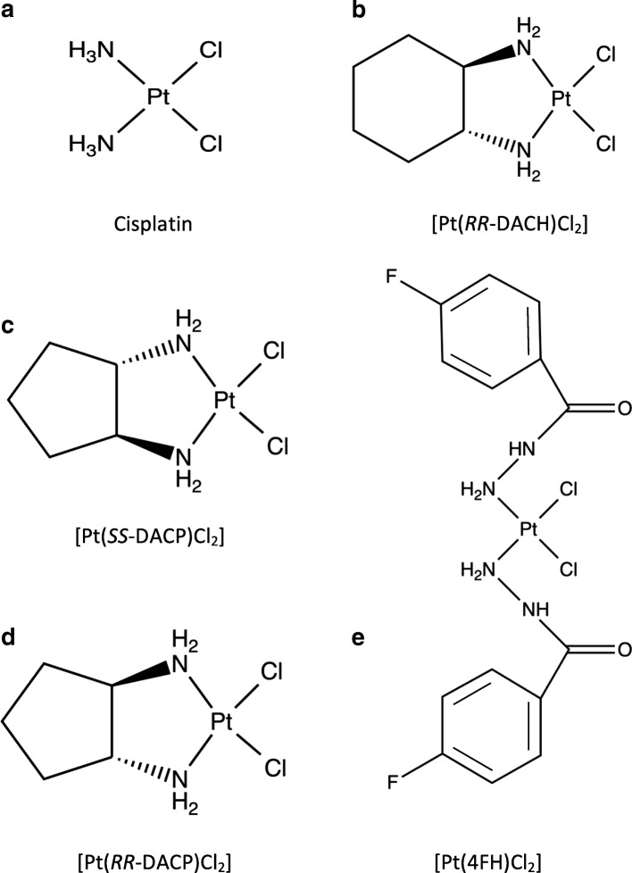
Background: Cisplatin has been widely used for the treatment of cancer and its antitumour activity is attributed to its capacity to form DNA adducts, predominantly at guanine residues, which impede cellular processes such as DNA replication and transcription. However, there are associated toxicity and drug resistance issues which plague its use. This has prompted the development and screening of a range of chemotherapeutic drug analogues towards improved efficacy. The biological properties of three novel platinum-based compounds consisting of varying cis-configured ligand groups, as well as a commercially supplied compound, were characterised in this study to determine their potential as anticancer agents.

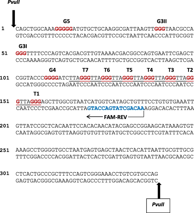
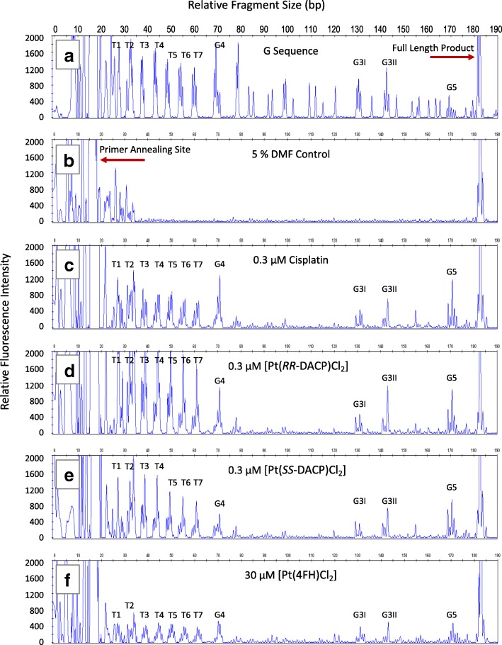
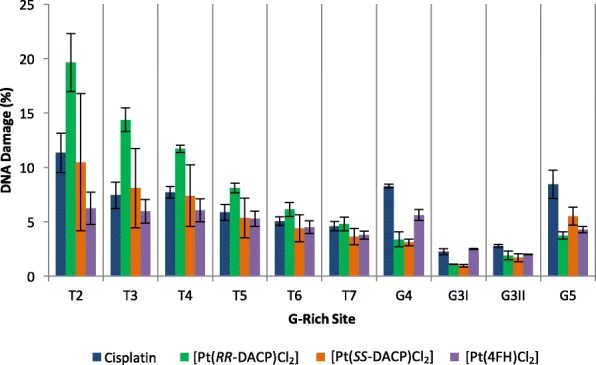
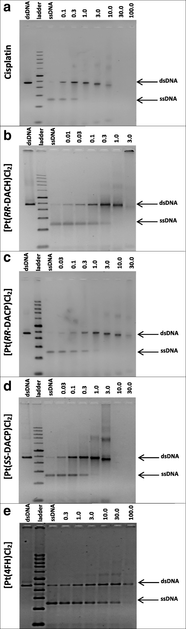
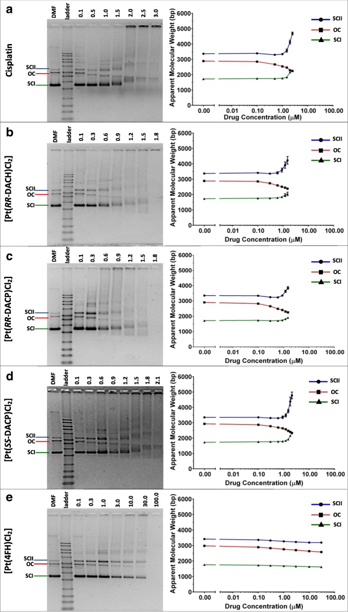
Methods: The linear amplification reaction was employed, in conjunction with capillary electrophoresis, to quantify the sequence specificity of DNA adducts induced by these compounds using a DNA template containing telomeric repeat sequences. Additionally, the DNA interstrand cross-linking and unwinding efficiency of these compounds were assessed through the application of denaturing and native agarose gel electrophoresis techniques, respectively. Their cytotoxicity was determined in HeLa cells using a colorimetric cell viability assay.

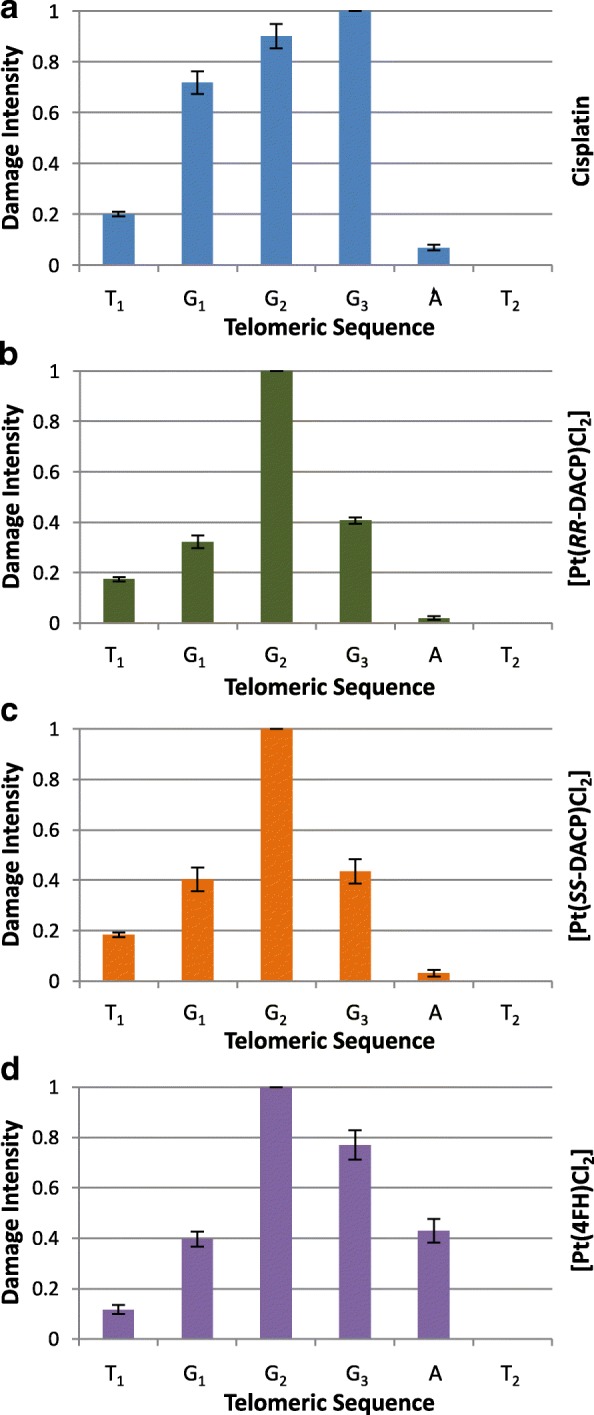
Results: All three novel platinum-based compounds were found to induce DNA adduct formation at the tandem telomeric repeat sequences. The sequence specificity profile at these sites was characterised and these were distinct from that of cisplatin. Two of these compounds with the enantiomeric 1,2-diaminocyclopentane ligand (SS and RR-DACP) were found to induce a greater degree of DNA unwinding than cisplatin, but exhibited marginally lower DNA cross-linking efficiencies. Furthermore, the RR-isomer was more cytotoxic in HeLa cells than cisplatin.

Conclusions: The biological characteristics of these compounds were assessed relative to cisplatin, and a variation in the sequence specificity and a greater capacity to induce DNA unwinding was observed. These compounds warrant further investigations towards developing more efficient chemotherapeutic drugs.

Keywords: Anticancer drug; Cisplatin; DNA adducts; Interstrand cross-linking; Linear amplification reaction; Sequence specificity; Telomeric repeat.


Conflict of interest statement

Ethics approval and consent to participate

Not applicable. However, we wish to acknowledge that in 1951 HeLa cells were taken without consent from Henrietta Lacks and we thank the Lacks family for their generous contributions to the biomedical community.

Consent for publication

Not applicable.

Competing interests

The authors declare that they have no competing interests.

Publisher’s Note

Springer Nature remains neutral with regard to jurisdictional claims in published maps and institutional affiliations.

Figures

Fig. 1
Fig. 1
The chemical structures of cisplatin, RR-DACH and the three novel platinum-based compounds used in this study. All compounds used in this study consist of a single platinum atom and two chloride atoms arranged in the cis configuration. Note that (a) cisplatin is a square planar compound containing two ammine groups attached to the central platinum atom. RR-DACH, contains an RR-configured cyclohexane ligand attached to two ammine groups. The novel compounds (c) [Pt(SS-DACP)Cl2] and (d) [Pt(RR-DACP)Cl2] consist of isomeric configured cyclopentane ligands. The novel compound (e) [Pt(4FH)Cl2] contains symmetrical fluorobenzoic hydrazide groups attached to ammine groups on either side of the platinum atom
Fig. 2
Fig. 2
Double-stranded sequence of the PvuII-cleaved pUC19/T7 DNA template. The upper strand of the sequence is written in the 5′ to 3′ direction. Sites of PvuII restriction enzyme cleavage are indicated by the bold vertical arrows and the annealing site of the FAM-REV primer is shown by a horizontal arrow, with the primer sequence highlighted in blue. The seven telomeric repeats sequences (T1 to T7) are underlined, with the guanines highlighted in red. Other sites of three or more consecutive guanines (G3I, G3II, G4 and G5) are highlighted in red
Fig. 3
Fig. 3
Electropherogram traces showing DNA damage induced by cisplatin and novel platinum-based compounds that was amplified by the LAR. a The G dideoxy sequencing lane used to determining the location of G-bases in the PvuII-cleaved pUC19/T7 sequence. b The 5% (v/v) DMF treated DNA, a negative control. DNA damage induced by (c) 0.3 μM cisplatin, (d) 0.3 μM [Pt(RR-DACP)Cl2] (e) 0.3 μM [Pt(SS-DACP)Cl2] and (f) 30 μM [Pt(4FH)Cl2]. The relative fluorescence intensity is plotted on the y-axis (left) and the DNA fragment size (bp) is plotted on the x-axis (top). The primer annealing site and full length extension product of 186 bp are highlighted. Damage peaks corresponding to sites of telomeric repeats T1 to T7 and the G-rich sites (G3I, G3II, G4 and G5) are labelled accordingly
Fig. 4
Fig. 4
Histograms showing the overall percentage of DNA damage induced by each compound in the PvuII-cleaved pUC19/T7 sequence. The percentage of overall DNA damage (y-axis) is shown for each G-rich site (x-axis). The overall DNA damage is the sum of damage at each individual base within the G-rich site. The error bars represent the SEM, determined from three separate experiments. The T1 site has been omitted as the DNA damage overlapped with artefact peaks present in the DMF control
Fig. 5
Fig. 5
Bar Charts showing average damage at individual bases of the telomeric repeats. The graphs for (a) cisplatin, (b) [Pt(RR-DACP)Cl2], (c) [Pt(SS-DACP)Cl2] and (d) [Pt(4FH)Cl2], are highlighted in blue, green, orange and purple, respectively. The damage intensity of each individual peak was normalised to a maximal value of 1. The error bars represent the SEM determined across each of three replicates at the T4, T5 and T6 TGGGAT sequences
Fig. 6
Fig. 6
Images of 1.2% (v/v) denaturing agarose gels showing ssDNA or compound-induced cross-linked dsDNA. Denaturing agarose gels showing the large (2364 bp) PvuII-cleaved fragment of pUC19 following treatment with (a) cisplatin, (b) [Pt(RR-DACH)Cl2], (c) [Pt(RR-DACP)Cl2], (d) [Pt(SS-DACP)Cl2], and (e) [Pt(4FH)Cl2]. DMF control samples of non-heat denatured (dsDNA) and heat denatured (ssDNA) plasmid are present on the left and right-hand side of the 1 kb ladder, respectively. Bands to the right correspond to heat-denatured samples that were prior treated with increasing concentration of compound (values are indicated in μM above each gel image)
Fig. 7
Fig. 7
Images of 1% (v/v) native agarose gels and their respective semi-log scatter plots, showing changes in the apparent molecular weight of the native pUC19 DNA conformations following treatment with (a) cisplatin, (b) [Pt(RR-DACH)Cl2], (c) [Pt(RR-DACP)Cl2], (d) [Pt(SS-DACP)Cl2] and (e) [Pt(4FH)Cl2]. Bands corresponding to the OC, SCI and SCII conformations of pUC19, are indicated on the left-hand side of each gel image. The DMF solvent control sample (pUC19 without drug treatment) is situated to the left of the 1 kb ladder. All other lanes contain DNA samples treated with increasing concentration of compound as indicated in μM above each gel image. The changes in apparent molecular weight of each pUC19 conformation is plotted on the semi-log scatter graphs to the right of each image. The band migration for the OC, SCI and SCII plasmid conformations are represented by red, green and blue lines, respectively. The error bars shown represent the SEM determined across three separate experiments

References

    1. Sedletska Y, Giraud-Panis MJ, Malinge JM. Cisplatin is a DNA-damaging antitumour compound triggering multifactorial biochemical responses in cancer cells: importance of apoptotic pathways. Curr Med Chem Anticancer Agents. 2005;5:251–265. doi: 10.2174/1568011053765967. - DOI - PubMed
    1. Eastman A. The formation, isolation and characterization of DNA adducts produced by anticancer platinum complexes. Pharmacol Ther. 1987;34:155–166. doi: 10.1016/0163-7258(87)90009-X. - DOI - PubMed
    1. Kelland L. The resurgence of platinum-based cancer chemotherapy. Nat Rev Cancer. 2007;7:573–584. doi: 10.1038/nrc2167. - DOI - PubMed
    1. Fichtinger-Schepman AMJ, Van der Veer JL, Den Hartog JHJ, Lohman PHM, Reedijk J. Adducts of the antitumor drug cis-diamminedichloroplatinum (II) with DNA: formation, identification, and quantitation. Biochemistry. 1985;24:707–713. doi: 10.1021/bi00324a025. - DOI - PubMed
    1. Jamieson ER, Lippard SJ. Structure, recognition, and processing of cisplatin- DNA adducts. Chem Rev. 1999;99:2467–2498. doi: 10.1021/cr980421n. - DOI - PubMed

LinkOut - more resources