. 2019 Jan 2;116(1):277-286.
doi: 10.1073/pnas.1811938116. Epub 2018 Dec 21.

PARL deficiency in mouse causes Complex III defects, coenzyme Q depletion, and Leigh-like syndrome

Affiliations

PARL deficiency in mouse causes Complex III defects, coenzyme Q depletion, and Leigh-like syndrome

Marco Spinazzi et al. Proc Natl Acad Sci U S A. .

Abstract

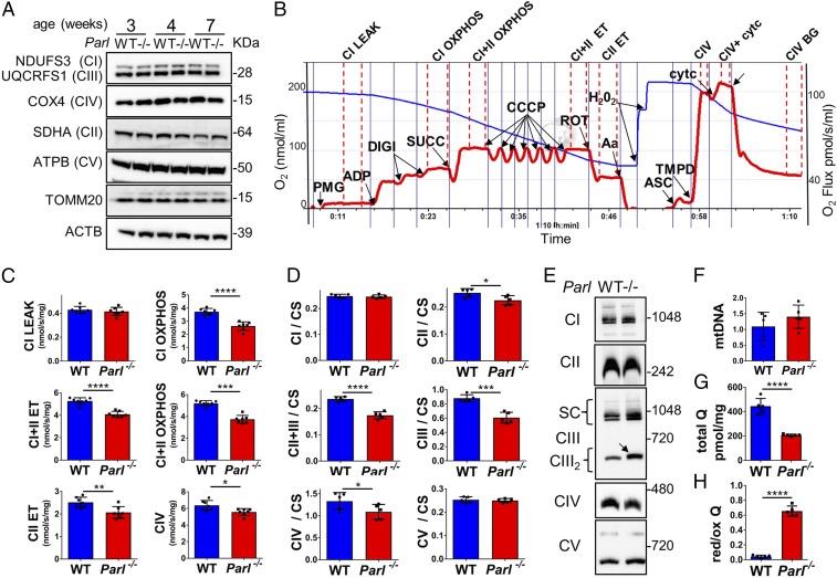
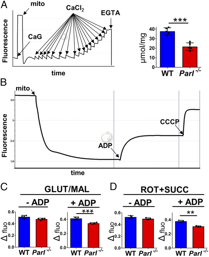
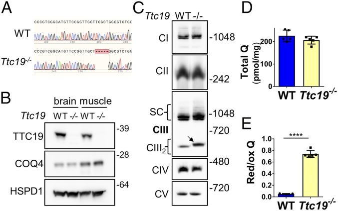
The mitochondrial intramembrane rhomboid protease PARL has been implicated in diverse functions in vitro, but its physiological role in vivo remains unclear. Here we show that Parl ablation in mouse causes a necrotizing encephalomyelopathy similar to Leigh syndrome, a mitochondrial disease characterized by disrupted energy production. Mice with conditional PARL deficiency in the nervous system, but not in muscle, develop a similar phenotype as germline Parl KOs, demonstrating the vital role of PARL in neurological homeostasis. Genetic modification of two major PARL substrates, PINK1 and PGAM5, do not modify this severe neurological phenotype. Parl-/- brain mitochondria are affected by progressive ultrastructural changes and by defects in Complex III (CIII) activity, coenzyme Q (CoQ) biosynthesis, and mitochondrial calcium metabolism. PARL is necessary for the stable expression of TTC19, which is required for CIII activity, and of COQ4, which is essential in CoQ biosynthesis. Thus, PARL plays a previously overlooked constitutive role in the maintenance of the respiratory chain in the nervous system, and its deficiency causes progressive mitochondrial dysfunction and structural abnormalities leading to neuronal necrosis and Leigh-like syndrome.

Keywords: Leigh syndrome; mitochondria; neurodegeneration; respiratory chain; rhomboid protease.


Conflict of interest statement

The authors declare no conflict of interest.

Figures

Fig. 1.
Fig. 1.
A Leigh-like encephalomyelopathy drives Parl/ phenotype. (A) Severe vacuolar neurodegeneration in a 7-wk-old Parl/ mouse brain (H&E staining; n ≥ 12). (Scale bar: 1,250 µm.) (Inset) Detail of the thalamus. (Scale bars: 100 µm.) (B) Severe neuronal loss in the gray matter of Parl/ lumbar spinal cord at 7 wk of age (RBFOX3 staining; n = 3 for WT, n = 6 for Parl/). (Scale bar: 125 µm.) (C) GFAP staining showing prominent astrogliosis in Parl/ medulla oblongata at 7 wk of age (n = 3 for WT, n = 6 for Parl/). (Scale bar: 125 µm.) (D) IBA1 staining in superior colliculus of the midbrain at 7 wk (n = 3 for WT, n = 6 for Parl/). (Scale bar: 125 µm.) (E) Combined Luxol fast blue and H&E staining show preservation of the white matter (stained in blue) in 7-wk-old Parl/ mice (n = 3 for WT, n = 7 for Parl/). (Scale bar: 750 µm.) (F) Parl in situ hybridization. DG, dentate gyrus; HY, hypothalamus; PC, pyriform cortex; TLM, thalamus. (Scale bar: 1 mm.) (Inset) Strong Parl expression in WT reticular neurons (magnification: 100×). (G) H&E stain of the inferior colliculus in the midbrain of a 7-wk-old Parl/ mouse showing vascular proliferation (n > 10). (Scale bar: 50 µm.) (H) Focal hemorrhage in the olivary nucleus of a 7-wk-old Parl/ mouse. Bilateral symmetrical hemorrhages have been detected in brainstems of four of seven Parl/ mice at 7 wk of age and in none of the WT littermates. (Scale bar: 250 µm.) (Inset) Bilateral symmetrical hemorrhages in medulla oblongata (arrows). (I) Survival curves of WT (n = 14), Parl/ (n = 14), ParlL/L::NesCre (n = 15), and ParlL/L::CkmmCre mice (n = 13). (J) Western blot analysis of PARL protein in brain, thymus, spleen, muscle, and liver mitochondria isolated from 7-wk-old WT and Parl/ and 13-wk-old ParlL/L::NesCre mice (n = 3). NDUFS3 is the loading control. (K) H&E stain of midbrains from 10-wk-old ParlL/L::NesCre (n = 4) and 7-wk-old Parl/ mice (n = 12). (Scale bar: 380 µm.) (LO) Severe atrophy of the skeletal muscle (L), liver (M), and spleen (N) but normal testis size (O) are seen in 11-wk-old ParlL/L::NesCre male mice compared with an age-matched WT control (n > 15). (P) H&E stain of thymus from WT (age 7 wk; n = 6), ParlL/L::NesCre (age 10–13 wk; n = 4), and Parl/ mice (age 7 wk; n = 12). ParlL/L::NesCre and Parl/ thymus are atrophic. (Scale bar: 200 µm.)
Fig. 2.
Fig. 2.
Parl/ neurodegeneration is preceded by mitochondrial structural changes and is characterized by necrosis. (A) EM images of medulla oblongata neuronal mitochondria of WT and Parl/ over time at 3, 5, and 7 wk of age. (Right) High-magnification inset of images at left. (Scale bar: 1 µm.) (B) Semithin section stained with toluidine blue shows vacuolization and disintegration of neurons (black arrowheads) in medulla oblongata of 7-wk-old Parl/ mouse compared with WT. (Scale bar: 50 µm.) (C) Representative EM images showing intracellular vacuolization (black arrowheads) in a 7-wk-old Parl/ thalamic neuron. (Scale bar: 5 µm.) (D) Permeability of mitochondrial outer membrane by ascorbate/TMPD-driven oxygen consumption rates in 6-wk-old WT and Parl/ purified free brain mitochondria (n = 7) before and after addition of 10 µM cytochrome c. Data represent average ± SD. (E) Western blot of WT and Parl/ brain cytosolic and mitochondrial fractions at 7 wk of age (n = 3) showing absent cytochrome c in the cytosol. Anti-AIF1 and anti-TUBA1A are the mitochondrial and cytosolic markers. cyt, purified cytosol; Hom, total homogenate; mito, purified mitochondria. (F) Immunoblot of 7-wk-old WT and Parl/ brain nuclei-enriched fractions with anti-PARP1 antibody. (G) Activated CASP3 staining shows absence of positive neurons in 7-wk Parl/ thalamus despite severe neurodegeneration (n = 4). (Scale bar: 100 µm.) (H) In the dentate gyrus, a brain area that does not degenerate in germline or in ParlL/L::NesCre mice, activated CASP3 staining shows scattered positive neurons (black arrows) to the same extent in WT and Parl/ mice (n = 4). (Scale bar: 50 µm.) (I) Cultured primary neurons were treated with 10 µM etoposide for 24 h and lysed. Total neuronal lysates were immunoblotted with anti-CASP3 and anti-PARP1 antibodies. The black arrows indicate the proteolyzed CASP3 and PARP1. ACTB is the loading control.
Fig. 3.
Fig. 3.
Pink1 and Pgam5 do not interact genetically with Parl deficiency in vivo. (A) Validation of Parl//Pgam5/, Parl//Pink1/, and Pink1//Pgam5/ double-KO mice and Parl//Pgam5//Pink1/ triple-KO mice. Brain mitochondria were isolated from mice of the indicated genotype and immunoblotted with PARL, PINK1, and PGAM5 antibodies. The white arrow indicates the mature form of PGAM5, the black arrow indicates the unprocessed form, and the gray arrow indicates alternatively processed form in Parl/ mitochondria. HSPD1 is the loading control. (B) Survival curves of Parl/ (n = 10), Pink1//Pgam5/ (n = 10), Parl//Pink1/ (n = 17), Parl//Pgam5/ (n = 16), and Parl//Pgam5//Pink1/ (n = 13). (C) H&E staining of midbrain coronal sections of 7-wk-old mice of the indicated genotypes (n > 3). (Scale bar: 500 µm.)
Fig. 4.
Fig. 4.
Defects in CIII and CoQ in Parl/ brain mitochondria. (A) Immunoblot of respiratory chain subunits (CI–CV), TOMM20, in WT and Parl/ whole brain lysates at 3, 4, and 7 wk of age (n = 4). ACTB is the loading control. (B) Representative trace illustrating the protocol for high-resolution respirometry in neuronal mitochondria. The blue trace indicates the O2 concentration and the red trace indicates its time derivative. Purified synaptosomes (50 µg) were loaded in Miro6 buffer. Digitonin (Digi) was titrated to achieve optimal synaptosomal permeabilization. Substrates are as follows: CI (PMG, pyruvate + malate + glutamate), CII (Succ, succinate), and CIV (ASC/TMPD, ascorbate + TMPD). The uncoupler is CCCP. Inhibitors are as follows: CI (ROT, rotenone), CIII (Aa, Antimycin a), and CIV (KCN, potassium cyanide). Respiratory states are indicated between red dashed lines. CI LEAK, CI-driven leak respiration; CI OXPHOS, CI-driven phosphorylating respiration; CI+II OXPHOS, phosphorylating respiration driven by combined activation of CI and II; CI+II ET, electron transfer capacity driven by combined CI and II; CII ET, ET driven by CII; CIV, CIV-driven respiration; Cytc, exogenous cytochrome c is added to evaluate the integrity of the outer mitochondrial membranes. H2O2 in the presence of catalase is used to reoxygenate the chamber. (C) Quantification of the respiratory states of permeabilized synaptosomes isolated from 6-wk-old WT and Parl/ mice (n = 7) as from the protocol in B. (D) Enzymatic activities of individual respiratory chain complexes and CII+CIII in brain mitochondria from 6-wk-old WT and Parl/ mice (n = 5) normalized to citrate synthase. (E) Blue native gel electrophoresis of purified brain mitochondria from 7-wk-old WT and Parl/ mice, followed by immunoblotting with anti-NDUFS3 (CI), anti-SDHA (CII), anti-UQCRFS1 (CIII), anti-COX4I1 (CIV), and anti-ATP5B (CV). The arrow indicates the upward mobility change of CIII2 in Parl/ (n = 3). (F) Mitochondrial DNA normalized by nuclear DNA in 7-wk-old WT and Parl/ brainstems (n = 5). (G) Concentration of total CoQ (Q9+Q10) measured by HPLC in brain tissue from 7-wk-old WT and Parl/ mice (n = 5). (H) CoQ red/ox ratio from the experiment in G. Bar graphs indicate average ± SD. Statistical significance calculated by two-sided Student t test: *P < 0.05, **P < 0.01, ***P < 0.001, and ****P < 0.0001.
Fig. 5.
Fig. 5.
Alterations in calcium uptake and membrane potential in Parl/ brain mitochondria. (A) Calcium-retaining capacity of purified brain mitochondria. (Left) Representative trace of a typical fluorimetric experiment using Calcium Green illustrates the protocol detailed in SI Appendix, Material and Methods. CaCl2, calcium chloride titrations; CaG, Calcium Green; EGTA, calcium chelator; Mito, mitochondria. (Right) Graph bars represent the quantifications of the maximal amount of exogenous calcium retained by WT and Parl/ brain mitochondria purified from 6-wk-old mice before observing calcium efflux (n = 5). (B) Mitochondrial membrane potential (i.e., Δψ) in brain mitochondria using safranin. A typical fluorimetric experiment illustrates the protocol detailed in SI Appendix, Material and Methods. CCCP is the uncoupler. Mito, mitochondria. (C and D) Quantifications of the experiments described in A using exactly 150 µg brain mitochondria from 7-wk-old WT and Parl/ mice (n = 5 in C, n = 4 in D). (C) Δψ using the CI substrates glutamate and malate (GLUT/MAL) without (leak state) and with ADP (phosphorylating state). (D) Δψ using the CII substrate succinate in presence of the CI inhibitor rotenone without and with ADP. The graph bars indicate the average ± SD. Statistical significances by two-sided t test: **P < 0.005 and ***P < 0.0005.
Fig. 6.
Fig. 6.
Restricted changes in the brain mitochondrial proteome induced by PARL deficiency explain the CIII and CoQ defects. (A) Volcano plot showing differentially regulated proteins in Parl/ brain mitochondria purified from 5-wk-old WT and Parl/ brains (n = 3) analyzed by MS. Significantly differentially regulated proteins are distributed outside the volcano cutoff of fold change >2 and P value <0.05. Differentially expressed mitochondrial proteins are plotted in red. Previously reported PARL substrates that did not reach statistical significance are plotted in black. The nonmitochondrial proteins TRABD, BCAP31, BCAN, EIF1A, and SETDB were not included in the graph because they appeared unchanged in the validation experiment in B. (B) Validation of the MS results and of previously reported PARL substrates PINK1, STARD7, CLPB, HTRA2, and OPA1. Brain mitochondria isolated from WT, Parl/, and ParlL/L::NesCre were analyzed by immunoblotting. White arrows indicate the mature (processed) form of the protein, black arrows indicate unprocessed forms, and gray arrows indicate alternatively processed forms in Parl/ mitochondria. The asterisk indicates bands of uncertain significance. (C) Time course of TTC19, CoQ biosynthesis proteins, and SQOR in WT and Parl/ brains. Total brain lysates from WT and Parl/ mice killed at 1, 3, 5, and 7 wk of age (n = 3) were analyzed by immunoblotting (original blots in SI Appendix, Fig. S8). The graph bars indicate the quantifications. Each protein was normalized with the loading control HSPD1 and expressed as percentage relative to the WT. The graph bars indicate the average ± SD. Statistical significances by two-sided t test: *P < 0.05, **P < 0.01, ***P < 0.001, and ****P < 0.0001.
Fig. 7.
Fig. 7.
TTC19 deficiency causes alterations in CIII and CoQ red/ox but not in CoQ concentration in the nervous system. (A) Generation of Ttc19/ mice by CRISPR/Cas9 technology. Ttc19/ mice have a 5-bp deletion in the first exon. (B) Immunoblot analysis of brain and muscle mitochondria with anti-TTC19 and COQ4 antibodies. HSPD1 is the loading control. (C) Blue native gel electrophoresis of purified brain mitochondria from 7-wk-old WT and Ttc19/ mice, followed by immunoblotting with anti-NDUFS3 (CI), anti-SDHA (CII), anti-UQCRFS1 (CIII), anti-COX4I1 (CIV), and anti-ATP5B (CV). The arrow indicates the upward mobility change of CIII2 in Ttc19/ mitochondria. (D) Concentration of total CoQ (Q9+Q10) in brains from 7-wk-old WT and Ttc19/ mice (n = 5). (E) CoQ red/ox ratio from the experiment in D. Bar graphs indicate average ± SD. Statistical significance calculated by two-sided Student t test: ****P < 0.0001.

References

    1. Spinazzi M, De Strooper B. PARL: The mitochondrial rhomboid protease. Semin Cell Dev Biol. 2016;60:19–28. - PubMed
    1. Düsterhöft S, Künzel U, Freeman M. Rhomboid proteases in human disease: Mechanisms and future prospects. Biochim Biophys Acta Mol Cell Res. 2017;1864:2200–2209. - PubMed
    1. Cipolat S, et al. Mitochondrial rhomboid PARL regulates cytochrome c release during apoptosis via OPA1-dependent cristae remodeling. Cell. 2006;126:163–175. - PubMed
    1. Anand R, et al. The i-AAA protease YME1L and OMA1 cleave OPA1 to balance mitochondrial fusion and fission. J Cell Biol. 2014;204:919–929. - PMC - PubMed
    1. Chao J-R, et al. Hax1-mediated processing of HtrA2 by Parl allows survival of lymphocytes and neurons. Nature. 2008;452:98–102. - PubMed

Publication types

MeSH terms