

Review
doi: 10.1042/BST20180316.
Epub 2018 Dec 21.
Transporter oligomerisation: roles in structure and function
Affiliations
- PMID: 30578344
- PMCID: PMC6393857
- DOI: 10.1042/BST20180316
Item in Clipboard
Review
Transporter oligomerisation: roles in structure and function
Biochem Soc Trans.
.
Abstract
Oligomerisation is a key feature of integral membrane transporters with roles in structure, function and stability. In this review, we cover some very recent advances in our understanding of how oligomerisation affects these key transporter features, with emphasis on a few groups of transporters, including the nucleobase ascorbate transporters, neurotransmitter sodium symporters and major facilitator superfamily members.
Keywords: function; membrane lipid; oligomerisation; protein structure; transporter.
© 2019 The Author(s).
Conflict of interest statement
The Authors declare that there are no competing interests associated with the manuscript.
Figures
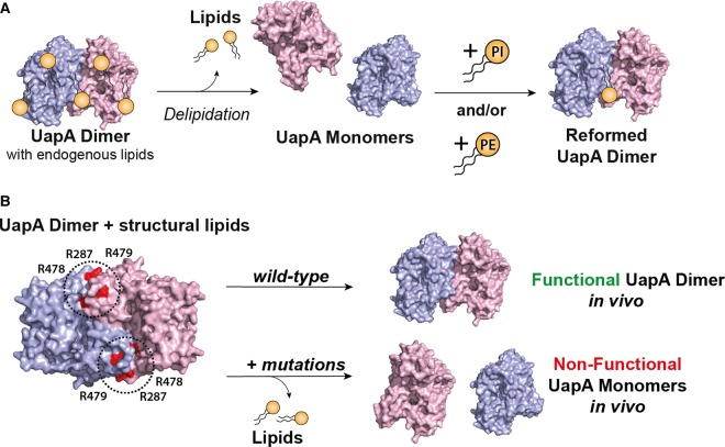
(A) UapA in detergent-based solution exists normally as a dimer in complex with membrane lipids as determined by native MS. Delipidation results in dissociation of the UapA dimers into the monomeric form. Dimers can be recovered by the addition of PI and PE both alone and in combination. (B) A potential lipid-binding site, comprising R287, R478 and R479, at the interface between the two protomers, was identified by molecular dynamics (MD) simulations. Mutation of all three Arg residues to Ala results in a loss of UapA transport function in the native host, Aspergillus nidulans. This triple mutant isolates almost exclusively in the monomeric form.
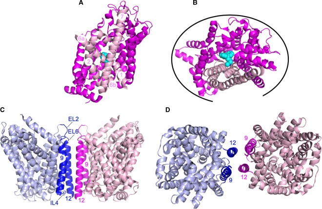
(A) DAT monomer (PDB: 4M48 [23]) looking through the membrane. TMHs 1, 2, 6 and 7 are coloured in pale pink with the remaining molecule coloured in magenta. The bound nortriptyline TCA, a tricyclic antidepressant, is shown in cyan. For clarity, some of the loop regions have been removed. (B) DAT monomer coloured and labelled as in (A) and shown from the extracellular side of the membrane. The black curve indicates all the regions predicted to be able form DAT:DAT interactions by MD simulations [29]. (C) LeuT dimer (PDB: 3TT1 [31]) looking through the membrane. One protomer is shown in pale blue and one in pale pink with the regions of the protomers (TMHs 9, 12 and extracellular loops 2 and 6 and intracellular loop 4 shown in dark blue and magenta). The different regions of the dimer interface are labelled. (D) LeuT dimer coloured and labelled as in (C) and shown from the extracellular side of the membrane.
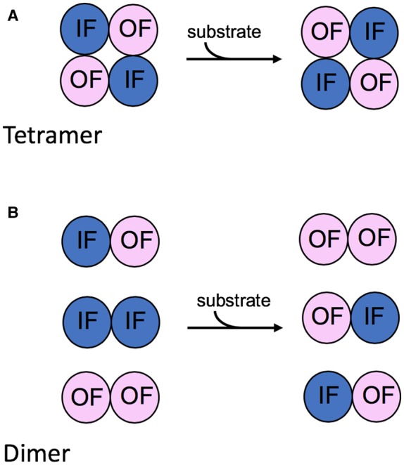
(A) In the tetrameric arrangement, there appears to be a concerted conformational rearrangement of the individual protomers. Substrate binding to the substrate-binding sites of two protomers exposed on either side of the membrane followed by transport results in the conformational rearrangement of all four protomers such that two protomers are always inward facing (IF) and two protomers are always outward facing (OF). (B) In contrast, in a dimer arrangement, the two protomers are associated but operate independently of one another.