. 2018 Dec 21;3(30):eaau8714.
doi: 10.1126/sciimmunol.aau8714.

Tuberculosis and impaired IL-23-dependent IFN-γ immunity in humans homozygous for a common TYK2 missense variant

Stéphanie Boisson-Dupuis  1   2   3 Noe Ramirez-Alejo  4 Zhi Li  5   6 Etienne Patin  7   8   9 Geetha Rao  10 Gaspard Kerner  2   3 Che Kang Lim  11   12 Dimitry N Krementsov  13 Nicholas Hernandez  4 Cindy S Ma  10   14 Qian Zhang  4   15 Janet Markle  4 Ruben Martinez-Barricarte  4 Kathryn Payne  10 Robert Fisch  4 Caroline Deswarte  2   3 Joshua Halpern  4 Matthieu Bouaziz  2   3 Jeanette Mulwa  4 Durga Sivanesan  16   17 Tomi Lazarov  18 Rodrigo Naves  19 Patricia Garcia  20 Yuval Itan  4   21   22 Bertrand Boisson  4   2   3 Alix Checchi  2   3 Fabienne Jabot-Hanin  2   3 Aurélie Cobat  2   3 Andrea Guennoun  15 Carolyn C Jackson  4   23 Sevgi Pekcan  24 Zafer Caliskaner  25 Jaime Inostroza  26 Beatriz Tavares Costa-Carvalho  27 Jose Antonio Tavares de Albuquerque  28 Humberto Garcia-Ortiz  29 Lorena Orozco  29 Tayfun Ozcelik  30 Ahmed Abid  31 Ismail Abderahmani Rhorfi  31   32 Hicham Souhi  31 Hicham Naji Amrani  31 Adil Zegmout  31 Frédéric Geissmann  18 Stephen W Michnick  16 Ingrid Muller-Fleckenstein  32 Bernhard Fleckenstein  32 Anne Puel  4   2   3 Michael J Ciancanelli  4 Nico Marr  15 Hassan Abolhassani  11   33 María Elvira Balcells  34 Antonio Condino-Neto  28 Alexis Strickler  35 Katia Abarca  36 Cory Teuscher  37 Hans D Ochs  38 Ismail Reisli  39 Esra H Sayar  39 Jamila El-Baghdadi  40 Jacinta Bustamante  4   2   3   41 Lennart Hammarström  11   12   42 Stuart G Tangye  10   14 Sandra Pellegrini  5   6 Lluis Quintana-Murci  7   8   9 Laurent Abel  4   2   3 Jean-Laurent Casanova  1   2   3   43   44
Affiliations

Tuberculosis and impaired IL-23-dependent IFN-γ immunity in humans homozygous for a common TYK2 missense variant

Stéphanie Boisson-Dupuis et al. Sci Immunol. .

Abstract

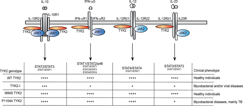
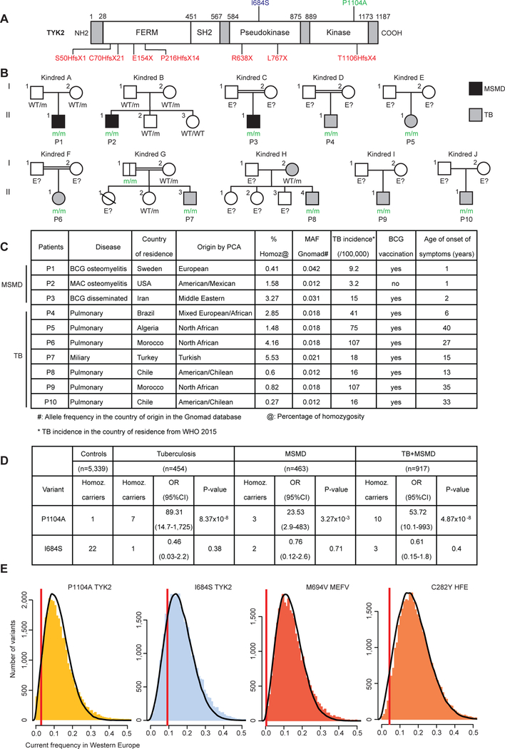
Inherited IL-12Rβ1 and TYK2 deficiencies impair both IL-12- and IL-23-dependent IFN-γ immunity and are rare monogenic causes of tuberculosis, each found in less than 1/600,000 individuals. We show that homozygosity for the common TYK2 P1104A allele, which is found in about 1/600 Europeans and between 1/1000 and 1/10,000 individuals in regions other than East Asia, is more frequent in a cohort of patients with tuberculosis from endemic areas than in ethnicity-adjusted controls (P = 8.37 × 10⁻⁸; odds ratio, 89.31; 95% CI, 14.7 to 1725). Moreover, the frequency of P1104A in Europeans has decreased, from about 9% to 4.2%, over the past 4000 years, consistent with purging of this variant by endemic tuberculosis. Surprisingly, we also show that TYK2 P1104A impairs cellular responses to IL-23, but not to IFN-α, IL-10, or even IL-12, which, like IL-23, induces IFN-γ via activation of TYK2 and JAK2. Moreover, TYK2 P1104A is properly docked on cytokine receptors and can be phosphorylated by the proximal JAK, but lacks catalytic activity. Last, we show that the catalytic activity of TYK2 is essential for IL-23, but not IL-12, responses in cells expressing wild-type JAK2. In contrast, the catalytic activity of JAK2 is redundant for both IL-12 and IL-23 responses, because the catalytically inactive P1057A JAK2, which is also docked and phosphorylated, rescues signaling in cells expressing wild-type TYK2. In conclusion, homozygosity for the catalytically inactive P1104A missense variant of TYK2 selectively disrupts the induction of IFN-γ by IL-23 and is a common monogenic etiology of tuberculosis.


Conflict of interest statement

Declaration of interest

The authors declare no competing interests.

Competing interests: The authors declare no competing interests.

Figures

Figure 1:
Figure 1:. Schematic representation of TYK2-dependent signaling pathways.
The cytokines, receptors, JAK and STAT complexes formed are indicated. A summary of the functionality of each pathway is provided for the various genotypes: WT TYK2, TYK2−/−, I684S and P1104A TYK2. ++++ means that the pathway is functional and optimal (corresponding to WT TYK2). + means that the function of the pathway is impaired but not completely abolished. The clinical phenotype of the individuals homozygous for the WT, P1104A and I684S alleles is indicated on the right.
Figure 2:
Figure 2:. Familial segregation and clinical information for patients homozygous for TYK2 P1104A.
(A) Schematic diagram of the TYK2 protein with its various domains (FERM, SH2, pseudokinase, and tyrosine kinase). The positions of the previously reported TYK2 mutations resulting in premature STOP codons are indicated in red. The positions of the I684S and P1104A polymorphisms are indicated in blue and green, respectively. (B) Pedigrees of the 10 TYK2-deficient families. Each generation is designated by a Roman numeral (I–II), and each individual by an Arabic numeral. The double lines connecting the parents indicate consanguinity based on interview and/or an homozygosity rate >4% estimated from the exome data. Solid shapes indicate disease status. Individuals whose genetic status could not be determined are indicated by “E?”, and “m” indicates a TYK2 P1104A allele. (C) Summary table of clinical details and origin of the patients associated with the minor allele frequency (MAF) in the country of origin. The incidence of tuberculosis (TB) in the country of residence (COR) is also mentioned. (D) Summary of WES, indicating the numbers of individuals with tuberculosis or MSMD and of controls carrying the I684S or P1104A variant of TYK2 in the homozygous state, and the associated p value and odds ratio. (E) Distributions of the current allele frequencies of variants that segregated 4,000 years ago at frequencies similar to those of the P1104A and I684S TYK2, M694V MEFV and C282Y HFE variants. The red vertical lines indicate the current frequency of the four variants of interest. Colored bars indicate the distribution of current allele frequency, in the 1,000 Genomes Project, for variants with frequencies in ancient European human DNA similar to those of the four candidate variants (52). Black lines indicate the distribution of simulated frequencies, in the present generation, for alleles with a past frequency similar to that of the four candidate variants, with propagation over 160 generations (corresponding to a period of ~4,000 years) under the Wright-Fisher neutral model. For instance, for the P1104A allele, which had a frequency ~9% in ancient Europeans, colored bars indicate the observed distribution of current frequencies for the 31,276 variants with a frequency of 8–10% 4,000 years ago; the black line indicates the distribution of frequencies for 100,000 simulated alleles obtained after 160 generations, under the Wright-Fisher neutral model.
Figure 3:
Figure 3:. Cellular responses to IFN-α, IL-12 and IL-23 in transduced EBV and HVS T cells.
TYK2-deficient EBV-B and HVS-T cells were transduced with a retrovirus generated with an empty vector (EV), or vectors encoding WT TYK2, or the P1104A, I684S or K930R TYK2 alleles. (A) Levels of TYK2 in transduced EBV-B (left) and HVS-T (right) cells, as determined by western blotting. (B) Levels of IL-12Rβ1 and IFN-αR1 in transduced EBV-B (left) and HVS-T (right) cells, as determined by flow cytometry. A p-value <0.001 in a two-tailed Student’s t test is indicated by ***. (C, D, F) Phosphorylation of JAKs and STATs in unstimulated (−) transduced EBV-B or HVS-T cells or in these cells following stimulation (+) with IFN-α (C) (pTYK2, pJAK1 and pSTAT1), IL-12 (D) (pTYK2, pJAK2, pSTAT1 and pSTAT4) and IL-23 (F) (pTYK2, pJAK2, pSTAT3 and pSTAT1), as assessed by western blotting with specific antibodies recognizing phospho-TYK2, phospho-JAK1, phospho-JAK2, phospho-STAT1, phospho-STAT4 and phospho-STAT3. (E) Phosphorylation of STAT4 in response to IFN-α and IL-12, as determined by flow cytometry in HVS-transduced T cells and expression as MFI. A p-value <0.01 and <0.001 in a two-tailed Student’s t test are indicated by ** and ***, respectively and ns means non significant. (G) IFN-β response of U1A (left) and MEF (right) cells, both lacking TYK2, after transduction with the indicated human and mouse TYK2 alleles, respectively, or with empty vector control (EV), as measured in an IFN-β-induced antiviral activity assay (see Materials and Methods). A unique dose is shown: 0.01ng/ml IFN-β for human cells and 1UI/ml for mouse cells.
Figure 4:
Figure 4:. Cellular responses to IFN-α, IL-12 and IL-23 in cell lines from patients
(A) TYK2 levels in EBV-B cells from two controls, two TYK2-deficient patients, two patients homozygous for TYK2 P1104A, two patients homozygous for TYK2 I684S and a patient compound heterozygous for the P1104A/I684S TYK2 alleles, as assessed by western blotting. (B) Levels of IL-12Rβ1 in EBV-B cells and HVS-T cells and of IFN-αR1 in EBV-B cells from controls, TYK2-deficient patients, patients homozygous for TYK2 P1104A, patients homozygous for TYK2 I684S and a patient compound heterozygous for P1104A/I684S TYK2 alleles, as assessed by flow cytometry. A p-value <0.01 in a two-tailed Student’s t test is indicated by **. ns = not significant. (C-E) Phosphorylation of JAKs and STATs in EBV-B or HVS-T cells of the indicated TYK2 genotypes, after stimulation with IFN-α (C) (pTYK2, pJAK1, pSTAT1 and pSTAT3), IL-12 (D) (pTYK2, pJAK2, and pSTAT4) or IL-23 (E) (pTYK2, pJAK2, and pSTAT3 ad pSTAT1), as determined by western blotting. (F) Phosphorylation of STAT3 after stimulation with IFN-α or IL-23, in HVS-T cells of the indicated genotypes, as assessed by flow cytometry. (G) Expression patterns on RNAseq of EBV-B cells stimulated with IFN-α. The heatmap represents the fold-change (FC) difference in expression before and after stimulation, on a log2 scale. Red blocks represent upregulated genes and blue blocks represent downregulated genes. The genes upregulated with a FC≥2.5, i.e. log2(FC) ≥1.3, in the group of controls are shown. (H) Relative levels of SOCS3 expression in EBV-B cells after IL-23 stimulation. (I) Percentage cell death for primary fibroblasts of the indicated genotype after VSV infection at various MOI, with and without IFN-β pretreatment.
Figure 5:
Figure 5:. Molecular mechanisms of impaired response to IL-23 by TYK2 P1104A
(A) In vitro kinase assay performed in the presence or absence of added ATP, on TYK2 immunopurified from human TYK2-deficient cells (U1A) stably reconstituted with either TYK2 WT or TYK2-P1104A. RecSTAT3 (upper panel) or recSTAT1 (lower panel) was added to the reaction mixture. The products of the reaction were analyzed by immunoblotting with Abs specific to the two activation loop tyrosine residues of TYK2 (Tyr1054–1055), phospho-STAT1 (Tyr701) or phospho-STAT3 (Tyr705). (B) Fold-change in RLU after stimulation with IL-23 (left) or IL-12 (right) in TYK2−/− cells stably reconstituted with IL-12Rβ1 and IL23R (left) or IL-12Rβ1 and IL-12Rβ2 (right). Cells were left untransfected, or were transfected with JAK2 and TYK2 fused to Rluc fragments, for the detection of interactions on PCA (RLU) of Rluc. The TYK2 used was either WT or P1104A. (C) Phosphorylation of JAK2, TYK2, STAT1 and STAT3 after stimulation with IL-12 and IL-23, in TYK2−/− and JAK2−/− fibrosarcoma cells reconstituted with IL-12Rβ1, IL-12Rβ2 and IL-23R. TYK2−/− cells were transfected with WT, P1104A or K930R TYK2 and JAK2−/− cells were transfected with WT, P1057A or K830E JAK2.
Figure 6:
Figure 6:. Analysis of primary cells from patients
(A) ELISA analysis of IFN-γ levels in whole blood after stimulation with BCG, or BCG plus IL-12, in controls, TYK2-deficient and IL-12Rβ1-deficient patients, and patients homozygous for the P1104A or I684S TYK2 alleles. p-values <0.05, <0.01, or <0.001 in two-tailed Student’s t tests are indicated by *, **, and ***, respectively. ns = not significant. (B) Production of IFN-γ from PBMCs stimulated with BCG, BCG plus IL-12, or BCG plus IL-23 in healthy controls, homozygous TYK2 P1104A patients, hyper-IgE patients with heterozygous STAT3 mutations (STAT3-DN), and patients with complete TYK2 and IL-12Rβ1 deficiencies, as determined by ELISA. (C) Percentages of IL-17A-, IL-17F- and IFN-γ positive CD4+ T cells after the stimulation of PBMCs from healthy controls and patients homozygous for TYK2 P1104A with PMA plus ionomycin. (D) In vitro differentiation of naïve CD4+ T cells from healthy controls, patients homozygous for P1104A TYK2 alleles and patients with TYK2 deficiency, following culture under Th17 (with IL-23) or Th1 (with IL-12) polarizing conditions, as determined by assessments of the induction of IL-17A/F and IFN-γ secretion, respectively. (E) Production of IFN-γ, IL-17A, IL-17F and IL-22 by naïve and memory CD4+ T cells from healthy controls, patients homozygous for P1104A TYK2, and TYK2-deficient patients, stimulated with TAE beads for 5 days.

References

    1. Houben RM, Dodd PJ, The Global Burden of Latent Tuberculosis Infection: A Re-estimation Using Mathematical Modelling. PLoS Med 13, e1002152 (2016). - PMC - PubMed
    1. Borgdorff MW et al., The incubation period distribution of tuberculosis estimated with a molecular epidemiological approach. Int J Epidemiol 40, 964–970 (2011). - PubMed
    1. Esmail H, Barry CE 3rd, Young DB, Wilkinson RJ, The ongoing challenge of latent tuberculosis. Philos Trans R Soc Lond B Biol Sci 369, 20130437 (2014). - PMC - PubMed
    1. WHO, Global tuberculosis report WHO/HTM/TB/2015.22, (2015).
    1. Abel L, El-Baghdadi J, Bousfiha AA, Casanova JL, Schurr E, Human genetics of tuberculosis: a long and winding road. Philos Trans R Soc Lond B Biol Sci 369, 20130428 (2014). - PMC - PubMed

Publication types