. 2019 Jan 15;92(3):e224-e233.
doi: 10.1212/WNL.0000000000006788. Epub 2018 Dec 21.

Word comprehension in temporal cortex and Wernicke area: A PPA perspective

Affiliations

Word comprehension in temporal cortex and Wernicke area: A PPA perspective

M-Marsel Mesulam et al. Neurology. .

Abstract

Objective: To explore atrophy-deficit correlations of word comprehension and repetition in temporoparietal cortices encompassing the Wernicke area, based on patients with primary progressive aphasia (PPA).

Methods: Cortical thickness in regions within and outside the classical Wernicke area, measured by FreeSurfer, was correlated with repetition and single word comprehension scores in 73 right-handed patients at mild to moderate stages of PPA.

Results: Atrophy in the Wernicke area was correlated with repetition (r = 0.42, p = 0.001) but not single word comprehension (r = -0.072, p = 0.553). Correlations with word comprehension were confined to more anterior parts of the temporal lobe, especially its anterior third (r = 0.60, p < 0.001). A single case with postmortem autopsy illustrated preservation of word comprehension but not repetition 6 months prior to death despite nearly 50% loss of cortical volume and severe neurofibrillary degeneration in core components of the Wernicke area.

Conclusions: Temporoparietal cortices containing the Wernicke area are critical for language repetition. Contrary to the formulations of classic aphasiology, their role in word and sentence comprehension is ancillary rather than critical. Thus, the Wernicke area is not sufficient to sustain word comprehension if the anterior temporal lobe is damaged. Traditional models of the role of the Wernicke area in comprehension are based almost entirely on patients with cerebrovascular lesions. Such lesions also cause deep white matter destruction and acute network diaschisis, whereas progressive neurodegenerative diseases associated with PPA do not. Conceptualizations of the Wernicke area that appear to conflict, therefore, can be reconciled by considering the hodologic and physiologic differences of the underlying lesions.


Figures

Figure 1
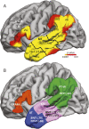
Figure 1. Atrophy–function correlations within 4 components of the language network
(A) Volume loss (mean [SD]) at false discovery rate ≤0.05 in the group of 73 patients with PPA compared to 38 neurologically healthy controls. Red and yellow patches show significant areas of volume loss. The numbers refer to mean changes in volume and SD. Significance is displayed as a p value. (B) Significant positive correlations between cortical thinning and language tasks. The numbers in parentheses indicate r values. AG = angular gyrus; AT = anterior temporal sector; BNT = Boston Naming Test; GRAM = % grammatically correct sentences in narrative; IFG = inferior frontal gyrus; IFG-B = inferior frontal gyrus, containing the Broca area; ITG = inferior temporal gyrus; MT = mid-temporal sector; MTG = middle temporal gyrus; PPVT = Peabody Picture Vocabulary Test; PT-W = parietotemporal sector encompassing the Wernicke area; REP = repetition task based on difficult items of the Western Aphasia Battery; SMG = supramarginal gyrus; STG = superior temporal gyrus; TP = temporal pole.
Figure 2
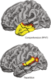
Figure 2. Atrophy–function correlations computed by individual vertex rather than by region of interest within all relevant temporoparietal cortices
Yellow and red areas show regions of significant correlations of cortical thinning (atrophy) with language comprehension and repetition scores. False discovery rate threshold was set at 0.05. AT = anterior temporal sector; MT = mid-temporal sector; MTG = middle temporal gyrus; PPVT = Peabody Picture Vocabulary Test; PT-W = parietotemporal sector encompassing the Wernicke area; STG = superior temporal gyrus.
Figure 3
Figure 3. Cortical thinning map in a patient with primary progressive aphasia (PPA) obtained 6 months before death
(A) Cortical thinning map for the patient with PPA obtained 6 months before death. False discovery rate ≤0.05. (B) Performance (expressed as percentage of perfect score) of the patient on language tasks 2.5 years before death (blue) and 6 months before death (red). (C) Stereologic tangle counts at autopsy in the right (purple) and left (tan) hemispheres. AG = angular gyrus; AT = anterior temporal sector; BNT = Boston Naming Test; ERC = entorhinal cortex; IFG = inferior frontal gyrus; IFG-B = inferior frontal gyrus, containing the Broca area; MT = mid-temporal sector; MTG = middle temporal gyrus; PPVT = Peabody Picture Vocabulary Test; pSTG = posterior superior temporal gyrus; PT-W = parietotemporal sector encompassing the Wernicke area; REP = repetition task based on difficult items of the Western Aphasia Battery; SMG = supramarginal gyrus; STG = superior temporal gyrus. Modified from Gefen et al.
Figure 4
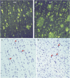
Figure 4. Alzheimer pathology in the patient with primary progressive aphasia (PPA)
(A, B) Thioflavin-S histofluorescence in layer 5 of the supramarginal gyrus (SMG) and posterior STG of the patient with PPA. Magnification ×200. (C) Cresylviolet stain of posterior superior temporal gyrus (STG) from the autopsy of a 54-year-old neurologically intact person shows pyramidal neurons (arrows) organized in columnar fashion. Magnification ×100. (D) Cresylviolet stain from posterior STG in the patient with PPA shows gliosis, angulated pyknotic neurons, and loss of columnar architecture. Magnification ×100.
Figure 5
Figure 5. Two models for reconciling lesion-symptom relationships of the Wernicke area in neurodegenerative vs cerebrovascular lesions
Double-headed arrows illustrate projection pathways. Red denotes destruction or severe functional loss; pink indicates partial functional disruption; green indicates sparing. The blue striped bars indicate destruction of the underlying projection pathway. AT = anterior temporal region; BR = Broca area; WER = Wernicke area.

References

    1. Bogen JE, Bogen GM. Wernicke's region: where is it? Ann NY Acad Sci 1976;280:834–843. - PubMed
    1. Wernicke C. Lehrbuch der Gehirnkrankheiten. Kassel: Theodore Fischer; 1881.
    1. Mesulam MM, Thompson CK, Weintraub S, Rogalski EJ. The Wernicke conundrum and the anatomy of language comprehension in primary progressive aphasia. Brain 2015;138:2423–2437. - PMC - PubMed
    1. Geschwind N Language and the brain. Sci Am 1972;226:76–83. - PubMed
    1. Binder JR. The Wernicke area. Neurology 2015;85:1–6. - PMC - PubMed

Publication types