. 2019 Feb;189(2):426-439.
doi: 10.1016/j.ajpath.2018.10.020. Epub 2018 Dec 21.

CCNA2 Ablation in Aged Mice Results in Abnormal rRNA Granule Accumulation in Hippocampus

Affiliations

CCNA2 Ablation in Aged Mice Results in Abnormal rRNA Granule Accumulation in Hippocampus

Michele J Alves et al. Am J Pathol. 2019 Feb.

Abstract

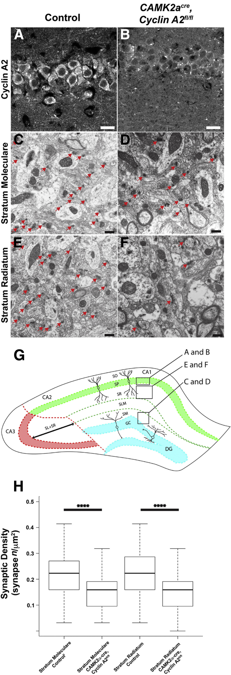
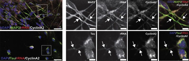
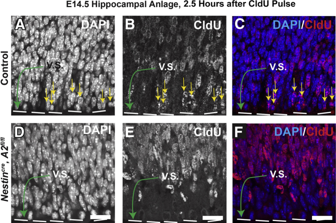
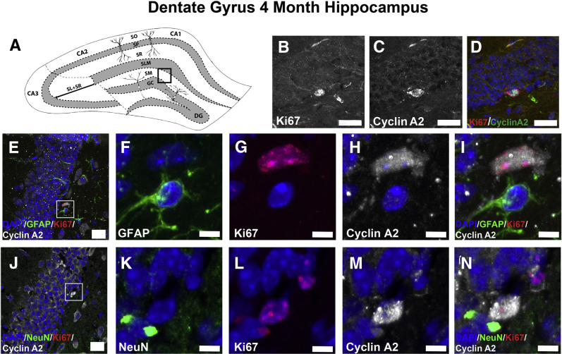
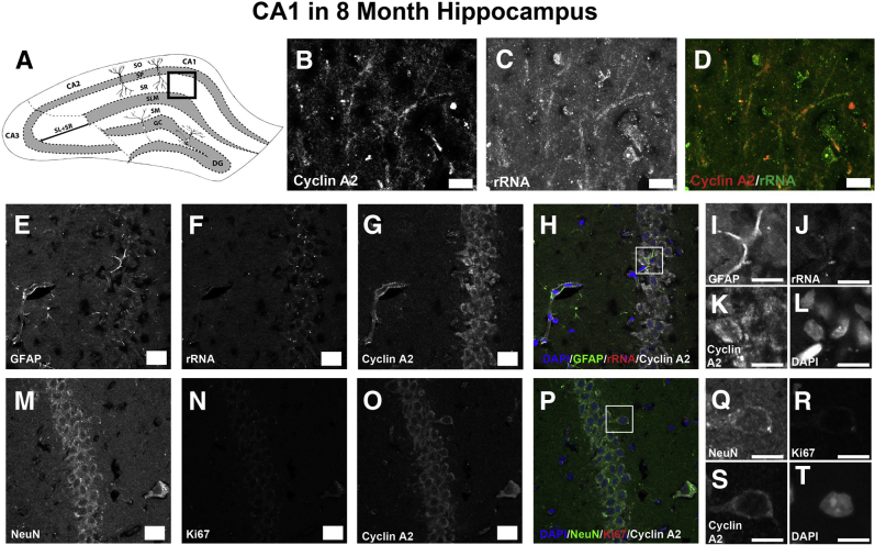
Mounting evidence in the literature suggests that RNA-RNA binding protein aggregations can disturb neuronal homeostasis and lead to symptoms associated with normal aging as well as dementia. The specific ablation of cyclin A2 in adult neurons results in neuronal polyribosome aggregations and learning and memory deficits. Detailed histologic and ultrastructural assays of aged mice revealed that post-mitotic hippocampal pyramidal neurons maintain cyclin A2 expression and that proliferative cells in the dentate subgranular zone express cyclin A2. Cyclin A2 loss early during neural development inhibited hippocampal development through canonical/cell-cycle mechanisms, including prolonged cell cycle timing in embryonic hippocampal progenitor cells. However, in mature neurons, cyclin A2 colocalized with dendritic rRNA. Cyclin A2 ablation in adult hippocampus resulted in decreased synaptic density in the hippocampus as well as in accumulation of rRNA granules in dendrite shafts. We conclude that cyclin A2 functions in a noncanonical/non-cell cycle regulatory role to maintain adult pyramidal neuron ribostasis.


Figures

Figure 1
Figure 1
Cyclin A2 loss in adult neurons results in decreased synaptic density in stratum moleculare (SM) and stratum radiatum (SR). A: Cyclin A2 expression in CA1 pyramidal neurons is lost in CamkIIacre, CCNA2fl/fl mice at 8 months. Genotypes are on top. A and B: Cyclin A2 immunostaining. Cyclin A2 shows cytoplasmic staining in CA1 pyramidal neurons, which is absent in the conditional cyclin A2 knockout. Perinuclear punctated staining present in B represents a non-specific reaction that is also present in A. C–F: Electron photomicrographs of control and mutant from the SM and SR layers of the hippocampus. Red arrows point to synapses. G: Illustration showing the anatomical hippocampal areas that are being analyzed with electron microscopy. H: Box-whisker plots of the synaptic density (per area) where the solid bar is the arithmetic median, the extent of the boxes is the interquartile range, and the whiskers are 1.5× the interquartile range. H: Results from P values derived from two-tailed homoscedastic t-test are plotted. All images and quantifications were obtained from 8-month–old mutant or control animals. ∗∗∗∗P < 0.0001. Scale bars: 10 μm (A and B); 500 nm (C–F). DG, dentate gyrus; GC, granule cell layer; SLM, stratum lacunosum-moleculare; SM, stratum moleculare; SO, stratum oriens; SP, stratum pyramidale; SR, stratum radiatum.
Figure 2
Figure 2
CamkIIacre, CCNA2fl/fl mice show no significant morphologic changes on Golgi impregnation relative to control mice. A–D: Examples of dentate granule neurons (DGNs) traced using Neurolucida obtained from Golgi impregnated slides. Genotype is indicated on top, and anatomical region and animal age are delineated on the left. A–G: Traced dentate granule neurons (A–D), dendrite complexity index (E), mean dendritic spine density (F), and complexity by Sholl analysis (G). No statistical difference is identified in neuronal complexity and dendritic spine density in dentate granule neurons between control and CamkIIacre, CCNA2fl/fl mice at 4 and 8 months. E–P: In whisker plots, the box of the whisker plot represents the interquartile range, the solid black bar represents the median, and the whiskers represent 1.5× the interquartile range.
Figure 3
Figure 3
Cyclin A2 colocalizes with rRNA and OligodT in hippocampal pyramidal neurons in vitro. Hippocampal pyramidal neuron cultures were generated and allowed to mature. These cells were then fixed and stained with anti–cyclin A2 (red) and anti-rRNA antibodies. A: A stitched tiled confocal image of a population of pyramidal neurons. The molecular markers are on the bottom left of each panel and color coded when appropriate. Small squares in A denote high-magnification images in B–D, E–G, and H–J. In these panels, each fluorescent channel is shown separately, followed by a merged channel. Yellow arrows indicate points where cyclin A2 and rRNA show colocalization, which tended to occur at locations of dendritic varicosities. Scale bars: 50 μm (A); 5 μm (B–J).
Figure 4
Figure 4
Cyclin A2 colocalizes with rRNA and OligodT in hippocampal pyramidal neurons in vitro. Hippocampal pyramidal neuron cultures were generated and allowed to mature. A–G: These cells were then fixed and stained with anti–cyclin D1 (green) and anti-rRNA antibodies. The molecular markers are on the bottom left of each panel and color coded when appropriate. A: A stitched tiled confocal image of a population of pyramidal neurons. A–G: Small squares in A denote high-magnification images in B–D and E–G. In these panels, each fluorescent channel is shown separately, followed by a merged channel. Red arrows point to rRNA accumulation on neuronal processes; green arrows, cyclin A2 accumulation; yellow arrows, points where cyclin A2 and rRNA show colocalization, which tends to occur at locations of dendritic varicosities. These accumulations tend to occur at separate locations on the neuronal process. Scale bars: 20 μm (A); 5 μm (B–G).
Figure 5
Figure 5
A: A confocal image of a pyramidal neuron in a mixed culture. The neuron is identified by its expression of MAP2 (green), the mRNA granules are identified by OligodT fluorescence in situ hybridization (red), and cyclin A2 is identified by staining in white. B–D: MAP2-positive processes maintain a light staining pattern for both OligodT and cyclin A2. E–G: OligodT is most highly enriched in the nucleus and perinuclear area. Scale bars: 20 μm (A); 10 μm (B–G).
Figure 6
Figure 6
Cyclin A2 is present in both axons and dendrites of hippocampal neurons. Cultured hippocampal neurons from postnatal day 0 mice were matured in vitro and immunostained for cyclin A2 and MAP2 or Tau to evaluate the colocalization of cyclin A2 with dendritic processes or axonal processes, respectively. Cyclin A2 immunostaining colocalizes to both MAP2-positive processes (arrows, top row) and Tau-positive processes (arrows, bottom row). Boxed areas are shown at higher magnification to the right. Scale bars: 20 μm (left column); 5 μm (higher magnification).
Figure 7
Figure 7
Cyclin A2 regulates proliferation of hippocampal progenitor cells. Brains from postnatal day 0 (P0) mice immunostained for cyclin A2 and Ki-67 to evaluate the colocalization of cyclin A2 in proliferative cells in the P0 dentate gyrus (A–E and K–O) and CA1 layer (F–J and P–T). All cyclin A2 immunoreactive cells colocalize to proliferative cells in the P0 hippocampus and do not colocalize to neurons or astrocytes. Scale bars: 20 μm (A, F, K, and P); 5 μm (B–E, G–J, L–O, and Q–T). GFAP, glial fibrillary acidic protein.
Figure 8
Figure 8
Cyclin A2 regulates proliferation of hippocampal progenitor cells. A–F: To test the effects of cyclin A2 ablation on proliferation in the embryonic day 14.5 (E14.5) hippocampal anlage, females impregnated with Nestincre, CCNA2fl/fl mice, and controls were pulsed with CldU for 2.5 hours. Green arrows denote the ventricular surface (V.S.); yellow arrows, progenitor cells that are CldU positive that have exited S-phase and have traversed to the ventricular surface via interkinetic nuclear migration. Scale bars = 10 μm (all images).
Figure 9
Figure 9
Cyclin A2 colocalizes to 4-month–old subgranular dentate gyrus progenitors. A: Hippocampal illustration denoting the localization of subsequently analyzed sections [boxed area indicates stratum moleculare (SM)]. B–D: Brain of control 4-month–old animal stained with anti–cyclin A2 and anti–Ki-67 antibodies. Note the presence of proliferative cells in the subgranular zone. E–I: Control dentate gyrus (DG) stained with glial fibrillary acidic protein (GFAP; green), Ki-67 (red), and cyclin A2 (white) shows no cyclin A2 colocalization to astrocytes in the DG. J–N: Control DG stained with NeuN (green), Ki-67 (red), and cyclin A2 (white) shows no cyclin A2 colocalization to neurons in the DG. Scale bars: 10 μm (B–D); 20 μm (E and J); 5 μm (F–I and K–N). DG, dentate gyrus; GC, granule cell layer; SLM, stratum lacunosum-moleculare; SM, stratum moleculare; SO, stratum oriens; SP, stratum pyramidale; SR, stratum radiatum.
Figure 10
Figure 10
Cyclin A2 colocalizes with rRNA in neurons of the 8-month–old hippocampal neuropil. A: Illustration depicting hippocampal area analyzed [boxed area indicates stratum radiatum (SR) of CA1]. B–D: The SR stained with rRNA and cyclin A2. Neurites show colocalization of cyclin A2 with rRNA in 8-month–old SR. E–L: Control SR stained with glial fibrillary acidic protein (GFAP; green), rRNA (red), and cyclin A2 (white) shows no cyclin A2 colocalization to astrocytes in the SR. Boxed area in H is shown at higher magnification in I–L. M–T: Control SR stained with NeuN (green), rRNA (red), and cyclin A2 (white) shows that cyclin A2 colocalizes to neurons in the 8-month–old SR. Boxed area in P is shown at higher magnification in Q–T. Scale bars: 10 μm (B–D, I–L, and Q–T); 20 μm (E–H and M–O). DG, dentate gyrus; GC, granule cell layer; SLM, stratum lacunosum-moleculare; SM, stratum moleculare; SO, stratum oriens; SP, stratum pyramidale; SR, stratum radiatum.
Figure 11
Figure 11
Cyclin A2 ablation in adult brains results in accumulation of rRNA in dendritic shafts in stratum radiatum (SR). A–D: Representative electron photomicrographs obtained from stratum moleculare (SM) and SR (designated to the left of each panel) in control or CamkIIacre, CCNA2fl/fl mice (denoted on the top). Significant accumulation of polyribosomes is noted by electron microscopy in dendritic shafts of 8-month–old mutants. Red arrows indicate the polyribosomes within a dendritic shaft. E: The polyribosome quantification is graphed. P values obtained from t-test are shown in the head-to-head comparisons. F and G: Representative low-power views of 4-month–old control or CamkIIacre, CCNA2fl/fl hippocampi, respectively. H–M: Representative low-power views of 8-month–old control or CamkIIacre, CCNA2fl/fl hippocampi, respectively (H and I). Boxed areas in H are seen at higher magnification in J and L. Boxed areas in I are seen at higher magnification in K and M. Note the presence of rRNA accumulation in both of these regions in the CamkIIacre, CCNA2fl/fl hippocampus (arrows). N and O: The quantification of rRNA immunostaining in the hippocampus. P < 0.05, ∗∗∗∗P < 0.0001. Scale bars: 500 nm (A–D); 50 μm (F–M).
Figure 12
Figure 12
rRNA accumulation in dendritic shafts of the stratum radiatum (SR) and stratum moleculare (SM) coincide with P-bodies. Panels show 8-month CamkIIacre, CCNA2fl/fl hippocampus immunostained for P-body marker GW182 (green) and rRNA (red) demonstrated on the SM and SR. Colocalization of GW182 with rRNA is denoted with arrows. Boxed areas are shown at higher magnification to the right. Scale bars: 20 μm (left column); 10 μm (higher magnification).

References

    1. Garcia-Moreno L.M., Conejo N.M., Pardo H.G., Gomez M., Martin F.R., Alonso M.J., Arias J.L. Hippocampal AgNOR activity after chronic alcohol consumption and alcohol deprivation in rats. Physiol Behav. 2001;72:115–121. - PubMed
    1. Pietrzak M., Rempala G., Nelson P.T., Zheng J.J., Hetman M. Epigenetic silencing of nucleolar rRNA genes in Alzheimer's disease. PLoS One. 2011;6:e22585. - PMC - PubMed
    1. Kiryk A., Sowodniok K., Kreiner G., Rodriguez-Parkitna J., Sonmez A., Gorkiewicz T., Bierhoff H., Wawrzyniak M., Janusz A.K., Liss B., Konopka W., Schutz G., Kaczmarek L., Parlato R. Impaired rRNA synthesis triggers homeostatic responses in hippocampal neurons. Front Cell Neurosci. 2013;7:207. - PMC - PubMed
    1. Tiku V., Jain C., Raz Y., Nakamura S., Heestand B., Liu W., Spath M., Suchiman H.E.D., Muller R.U., Slagboom P.E., Partridge L., Antebi A. Small nucleoli are a cellular hallmark of longevity. Nat Commun. 2016;8:16083. - PMC - PubMed
    1. Ramaswami M., Taylor J.P., Parker R. Altered ribostasis: RNA-protein granules in degenerative disorders. Cell. 2013;154:727–736. - PMC - PubMed

Publication types

MeSH terms