Review
. 2019 Mar:92:5-13.
doi: 10.1016/j.yebeh.2018.11.031. Epub 2018 Dec 21.

Driving status of patients with generalized spike-wave on EEG but no clinical seizures

Affiliations
Review

Driving status of patients with generalized spike-wave on EEG but no clinical seizures

Prince Antwi et al. Epilepsy Behav. 2019 Mar.

Abstract

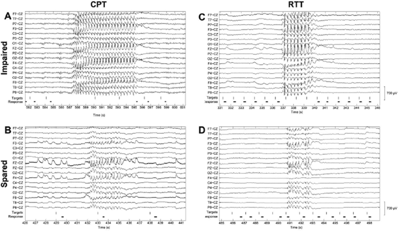
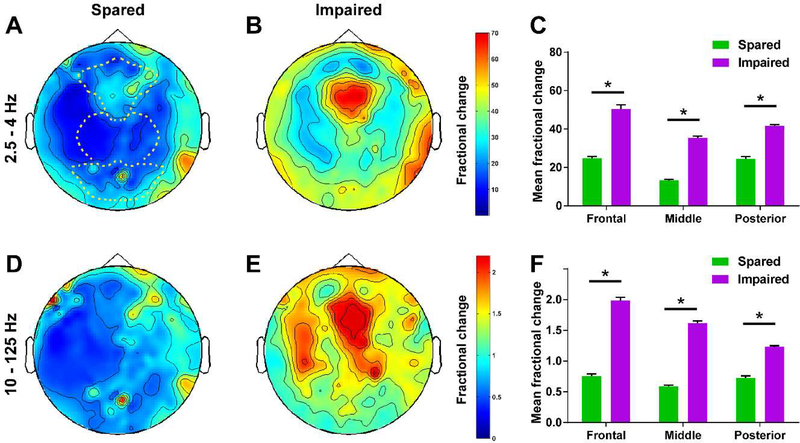
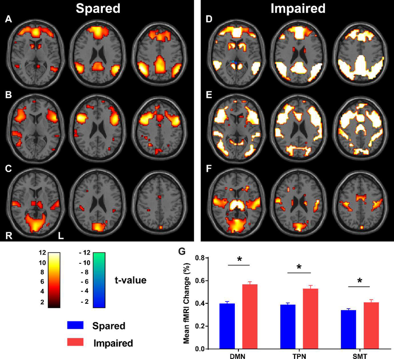
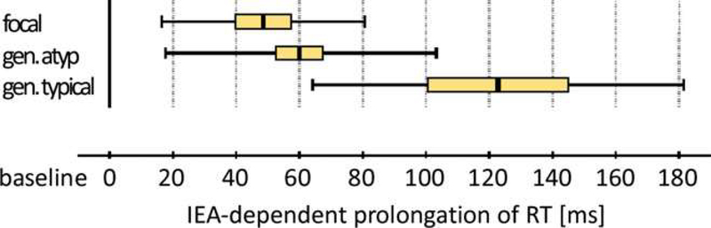
Generalized spike-wave discharges (SWDs) are the hallmark of generalized epilepsy on the electroencephalogram (EEG). In clinically obvious cases, generalized SWDs produce myoclonic, atonic/tonic, or absence seizures with brief episodes of staring and behavioral unresponsiveness. However, some generalized SWDs have no obvious behavioral effects. A serious challenge arises when patients with no clinical seizures request driving privileges and licensure, yet their EEG shows generalized SWD. Specialized behavioral testing has demonstrated prolonged reaction times or missed responses during SWD, which may present a driving hazard even when patients or family members do not notice any deficits. On the other hand, some SWDs are truly asymptomatic in which case driving privileges should not be restricted. Clinicians often decide on driving privileges based on SWD duration or other EEG features. However, there are currently no empirically-validated guidelines for distinguishing generalized SWDs that are "safe" versus "unsafe" for driving. Here, we review the clinical presentation of generalized SWD and recent work investigating mechanisms of behavioral impairment during SWD with implications for driving safety. As a future approach, computational analysis of large sets of EEG data during simulated driving utilizing machine learning could lead to powerful methods to classify generalized SWD as safe vs. unsafe. This may ultimately provide more objective EEG criteria to guide decisions on driving safety in people with epilepsy.

Keywords: Absence seizures; Consciousness; Driving safety; Driving simulation; Epilepsy; Subclinical epileptiform discharges.


Conflict of interest statement

Conflicts of Interest

None of the authors has any conflict of interest to disclose regarding this article.

Figures

Figure 1.
Figure 1.. Examples of EEG recordings showing generalized spike-wave discharges with impaired versus spared performance.
On both the continuous performance task (CPT, A and B) and repetitive tapping task (RTT, C and D), generalized SWDs that resulted in impaired performance (A and C) lasted longer than those that spared performance (B and D). Shown here are out-of-scanner high-density EEG recordings with a limited number of channels shown for ease of viewing. Vertical lines indicate presentation of target letters during behavioral tasks. For CPT (A and B), the targets consisted of the letter “X” presented in a stream of other letters appearing once each second. For RTT (C and D), the targets consisted of any letter presented once each second. Durations of button presses are indicated by the heavy black horizontal lines. Reproduced with permission from Guo et al., Impaired consciousness in patients with absence seizures investigated by functional MRI, EEG, and behavioural measures: a cross-sectional study. Lancet Neurology, 2016; 15(13) 1336–1345.
Figure 2.
Figure 2.. Generalized SWDs with impaired performance on CPT and RTT are associated with greater EEG amplitude in widespread brain regions.
(A and B) Wave component of SWDs represented by head maps of 256 channel high-density EEG power in the 2.5–4Hz frequency range for both performance sparing (A) and performance-impairing (B) SWDs. (C) Mean fractional EEG power in the 2.5–4Hz frequency range for seizures with spared versus impaired performance. (D and E) Spike component of SWDs represented by head maps of EEG power in the 10–125Hz frequency range for both spared (D) or impaired (E) performance. (F) Mean fractional EEG power in the 10–125Hz frequency range for seizures with spared versus impaired performance. Color scale bars are EEG power during seizures divided by baseline power before seizures (fractional power). The top color bar is for panels (A) and (B), and the bottom bar is for (D) and (E). Dashed lines in in (A) show regions used for analysis in (C) and (F) (frontal, middle, and posterior EEG contacts). *p<0.0001. Error bars represent standard error. Analysis is based on data from 30 performance-sparing seizures in 5 patients and 26 performance-impairing seizures in 8 patients. Reproduced with permission from Guo et al., Impaired consciousness in patients with absence seizures investigated by functional MRI, EEG, and behavioural measures: a cross-sectional study. Lancet Neurology, 2016; 15(13) 1336–1345.
Figure 3.
Figure 3.. fMRI signals in three established brain networks during generalized spike-wave seizures that spared or impaired performance on behavior tests.
(A, B and C) Axial brain t-maps with fMRI signals for generalized SWDs with spared performance in the default-mode network (DMN) (A), task-positive network (TPN) (B), and primary sensorimotor-thalamic network (SMT) (C). (D, E and F) Corresponding axial brain t-maps with fMRI signals for generalized SWDs with impaired performance. Color scale bars show t values. Hot colors (white-orange) indicate significant fMRI changes in the same direction as the network-specific hemodynamic response functions. Cool colors (green-blue) indicate changes in the opposite direction. (G) Mean percentage change in BOLD-fMRI signal across seizures in each network. Analysis is based on data from 93 performance-sparing seizures in 17 patients and 112 performance-impairing seizures in 22 patients. Reproduced with permission from Guo et al., Impaired consciousness in patients with absence seizures investigated by functional MRI, EEG, and behavioural measures: a cross-sectional study. Lancet Neurology, 2016; 15(13) 1336–1345.
Figure 4.
Figure 4.. Typical generalized spike-wave discharges prolong reaction time more than focal and atypical generalized spike-wave discharges on a computerized driving game.
Reaction times during interictal epileptiform activity (IEA) including focal, atypical and typical generalized spike-wave discharges are shown. IEA resulting in lapses or crashes during testing are excluded in calculating the depicted reaction times. Center value = mean, colored box = standard error of the mean, range bar = standard deviation. Reproduced with permission from Nirkko et al., Virtual car accidents of epilepsy patients, interictal epileptic activity, and medication. Epilepsia, 2016, 57(5) 832–840.

References

    1. Blumenfeld H. Cellular and Network Mechanisms of Spike-Wave Seizures. Epilepsia 2005;46: 21–33. - PubMed
    1. Ebersole JS, Pedley TA. Current Practice of Clinical Electroencephalography, 3rd Edition. Philadelphia, PA: Lippincott Williams & Wilkins; 2003.
    1. Scheffer IE, Berkovic S, Capovilla G, Connolly MB, French J, Guilhoto L, Hirsch E, Jain S, Mathern GW, Moshe SL, Nordli DR, Perucca E, Tomson T, Wiebe S, Zhang YH, Zuberi SM. ILAE classification of the epilepsies: Position paper of the ILAE Commission for Classification and Terminology. Epilepsia 2017;58: 512–521. - PMC - PubMed
    1. Scheffer IE, Berg AT. Classification and clinical features of absence epilepsies: how evidence leads to changing concepts. Epilepsia 2008;49: 2140–1. - PubMed
    1. Penry JK, Porter RJ, Dreifuss RE. Simultaneous recording of absence seizures with video tape and electroencephalography. A study of 374 seizures in 48 patients. Brain 1975;98: 427–40. - PubMed

Publication types