Review
. 2018 Dec;178(4):432-439.
doi: 10.1002/ajmg.c.31666.

Rhombencephalosynapsis: Fused cerebellum, confused geneticists

Affiliations
Review

Rhombencephalosynapsis: Fused cerebellum, confused geneticists

Kimberly A Aldinger et al. Am J Med Genet C Semin Med Genet. 2018 Dec.

Abstract

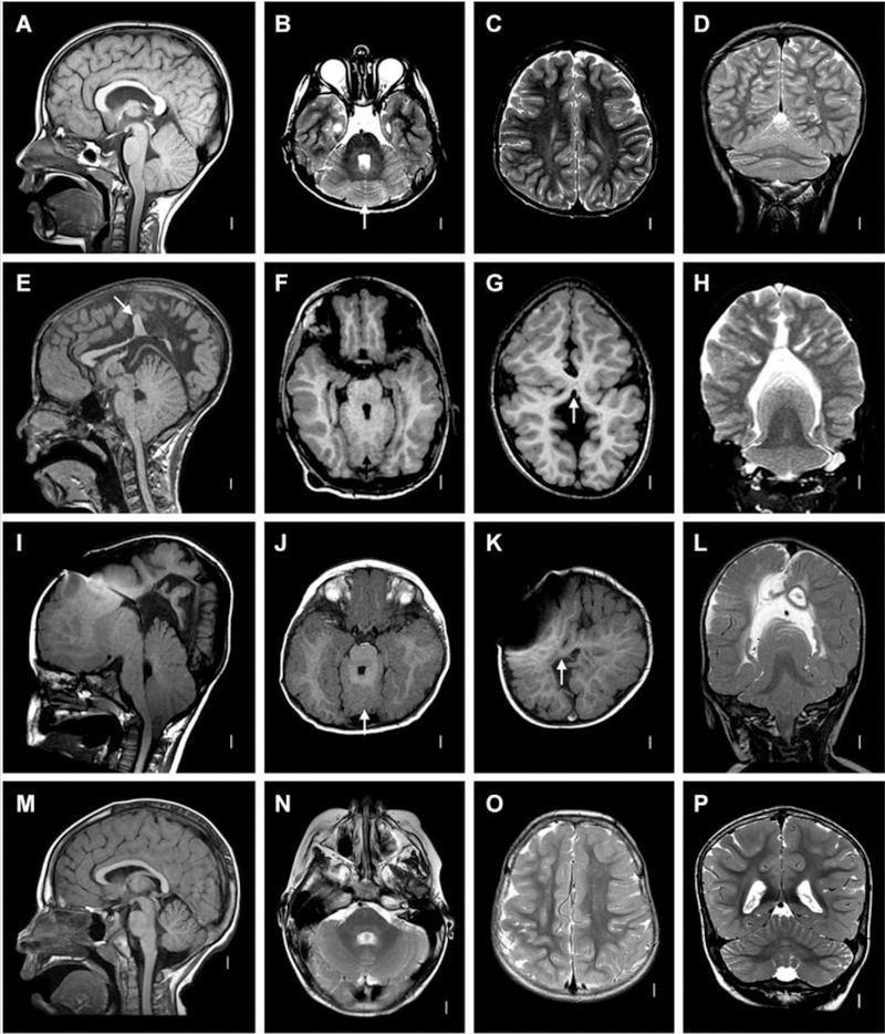
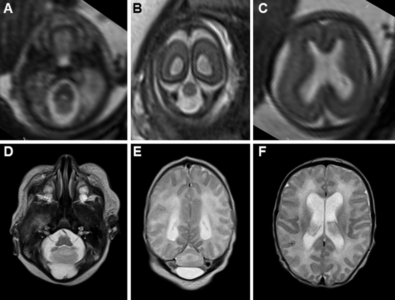
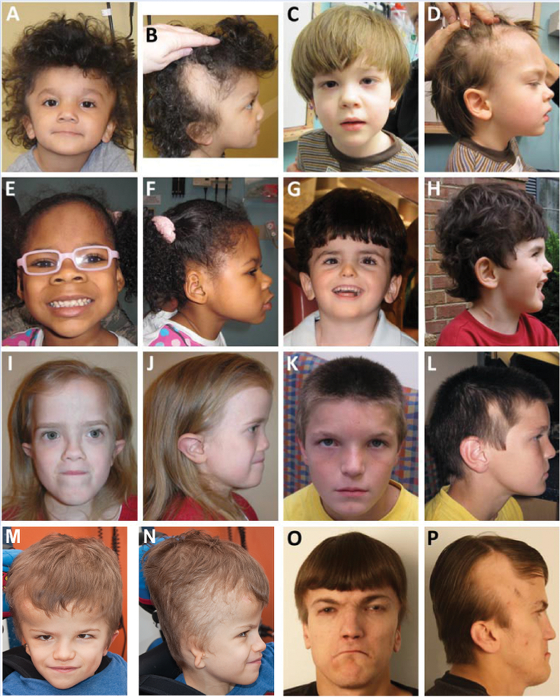
Rhombencephalosynapsis (RES) is a unique cerebellar malformation characterized by fusion of the cerebellar hemispheres with partial or complete absence of a recognizable cerebellar vermis. Subsets of patients also have other brain malformations such as midbrain fusion with aqueductal stenosis, characteristic craniofacial features (prominent forehead, flat midface, hypertelorism, ear abnormalities), and somatic malformations (heart, kidney, spine, and limb defects). Similar to known genetic brain malformations, the RES cerebellar malformation is highly stereotyped, yet no genetic causes have been identified. Here, we outline our current understanding of the genetic basis for RES, discuss limitations, and outline future approaches to identifying the causes of this fascinating brain malformation.

Keywords: RES; cerebellum; genetics; neuroimaging.


Figures

FIGURE 1
FIGURE 1
Postnatal radiographic features of RES. A–D: Patient 48. Midline sagittal T1-weighted image through the cerebellum demonstrates hemispheric rather than vermian configuration (A). Axial T2 through the cerebellum shows fusion of white matter across the midline and a keyhole-shaped 4th ventricle (B). Axial T2 through though the cerebral hemispheres shows normal anatomy (C). Coronal T2 showing continuity of cerebellar folia across the midline without an intervening vermis (D). E–H: Patient 46. Midline sagittal T1-weighted image demonstrating a towering cerebellum with hemispheric architecture. Note the absence of a visible aqueduct. This patient had severe congenital hydrocephalus. Many of the supratentorial abnormalities (arrow) are likely a consequence of distortion from hydrocephalus and subsequent decompression (E). Axial T1 through the cerebellum demonstrating similar findings as the patient above (F). Axial T1 through the cerebral hemispheres shows an area of white matter continuity suspicious for mild HPE, but likely representing post-hydrocephalus distortion (G). Coronal T2 showing a towering cerebellum with upward displacement through the tentorial notch (H). I–L: Patient 14. I, Mid-sagittal and axial T1 demonstrating similar findings to Patient 46. This patient also had severe congenital hydrocephalus (J). Axial T1 showing area suspicious for HPE (arrow), though post-hydrocephalic distortion makes this difficult to confirm (K). Coronal T2 demonstrating similar findings to Patient 46 (L). M–P: normal brain. Reproduced from Tully, et al. (2012) with permission.
FIGURE 2
FIGURE 2
Pre- and post-natal brain MRI features of RES. (A–C) Fetal MRI at 21 and 4/7 weeks gestational age demonstrates severe cerebellar hypoplasia (A and B) and mild ventriculomegaly (C). (D-F) Neonatal MRI of the same patient demonstrates severe cerebellar hypoplasia (D and E) and mild ventriculomegaly (F) in the same patient.
FIGURE 3
FIGURE 3
Faces of patients with RES showing typical facial features including hypertelorism (A, G, I K, L), prominent forehead (all), flat midface/midface retrusion (all except D), alopecia (B, D, L, P), turricephaly (N and P), and ear abnormalities (low, position (B, J, P), posterior rotation (B, D, L, P), and microtia (N)). Adapted from Tully, et al. (2012) with permission.

References

    1. Accogli A, & Srour M (2018). Teaching Video NeuroImages: Figure 8 head-shaking stereotypy in rhombencephalosynapsis. Neurology, 90(20), e1832–e1833. doi:10.1212/WNL.0000000000005531 - DOI - PubMed
    1. Barth PG (2008). Rhombencephalosynapsis. Handb Clin Neurol, 87, 53–65. doi:10.1016/S0072-9752(07)87004-7 - DOI - PubMed
    1. Chemli J, Abroug M, Tlili K, & Harbi A (2007). Rhombencephalosynapsis diagnosed in childhood: clinical and MRI findings. Eur J Paediatr Neurol, 11(1), 35–38. doi:10.1016/j.ejpn.2006.09.007 - DOI - PubMed
    1. Chong JX, Buckingham KJ, Jhangiani SN, Boehm C, Sobreira N, Smith JD, . . . Bamshad MJ (2015). The Genetic Basis of Mendelian Phenotypes: Discoveries, Challenges, and Opportunities. Am J Hum Genet, 97(2), 199–215. doi:10.1016/j.ajhg.2015.06.009 - DOI - PMC - PubMed
    1. de Mattos VF, Graziadio C, Machado Rosa RF, Lenhardt R, Alves RP, Trevisan P, . . . Zen PR (2014). Gomez-Lopez-Hernandez syndrome in a child born to consanguineous parents: new evidence for an autosomal-recessive pattern of inheritance? Pediatr Neurol, 50(6), 612–615. doi:10.1016/j.pediatrneurol.2014.01.035 - DOI - PubMed

Publication types

MeSH terms