

Higher Pulsatility in Cerebral Perforating Arteries in Patients With Small Vessel Disease Related Stroke, a 7T MRI Study
- PMID: 30580730
- PMCID: PMC6314503
- DOI: 10.1161/STROKEAHA.118.022516
Higher Pulsatility in Cerebral Perforating Arteries in Patients With Small Vessel Disease Related Stroke, a 7T MRI Study
Abstract
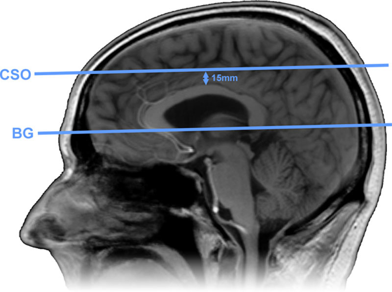
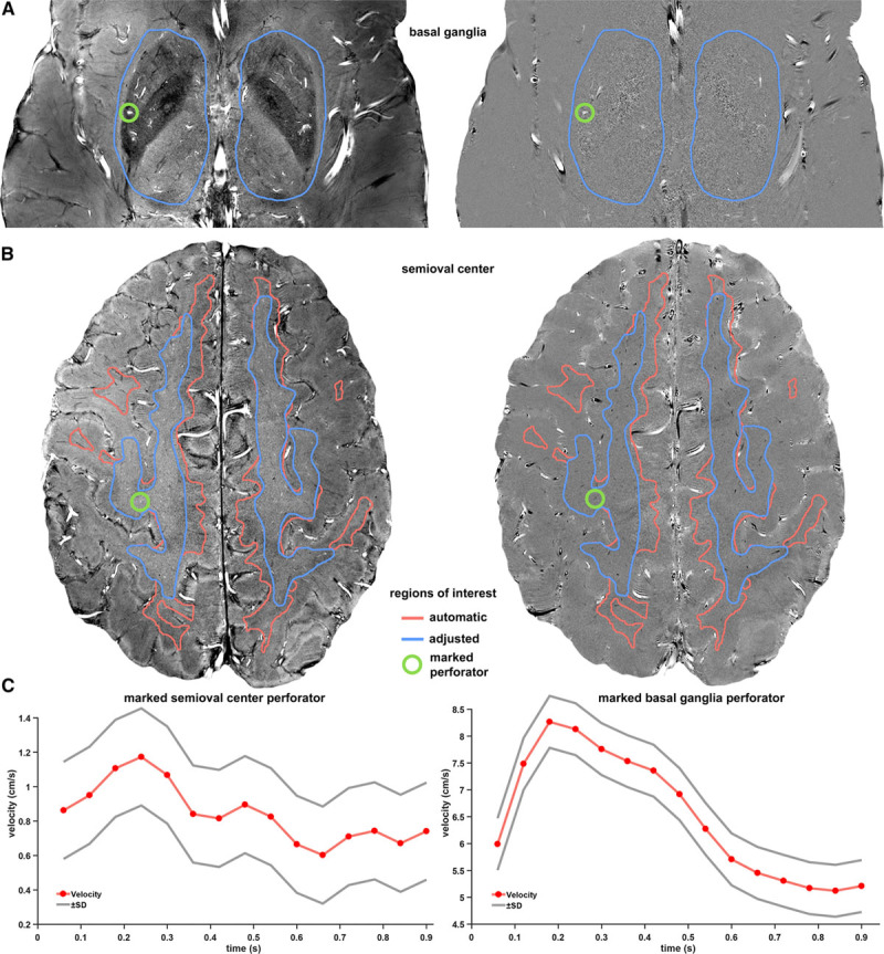
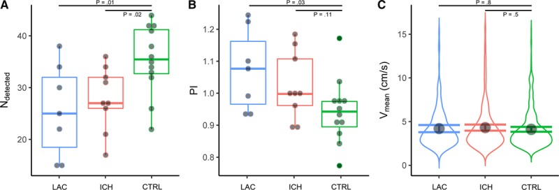
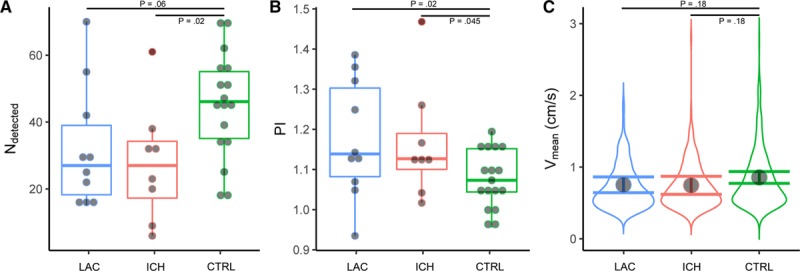
Background and Purpose- Cerebral small vessel disease (SVD) is a major cause of stroke and dementia, but underlying disease mechanisms are still largely unknown, partly because of the difficulty in assessing small vessel function in vivo. We developed a method to measure blood flow velocity pulsatility in perforating arteries in the basal ganglia and semioval center. We aimed to determine whether this novel method could detect functional abnormalities at the level of the small vessels in patients with stroke attributable to SVD. Methods- We investigated 10 patients with lacunar infarction (mean age 61 years, 80% men), 11 patients with deep intracerebral hemorrhage (ICH) considered to be caused by SVD (ICH, mean age 58 years, 82% men) and 18 healthy controls that were age- and sex-matched. We performed 2-dimensional phase contrast magnetic resonance imaging at 7 T to measure time-resolved blood flow velocity in cerebral perforating arteries of the semioval center and the basal ganglia. We compared the number of detected arteries, pulsatility index and mean velocity between the patient groups and controls. Results- In the basal ganglia, the number of detected perforators was lower in lacunar infarction (26±9, P=0.01) and deep ICH patients (28±6, P=0.02) than in controls (35±7). The pulsatility index in the basal ganglia was higher in lacunar infarction (1.07±0.13, P=0.03), and deep ICH patients (1.02±0.11, P=0.11), than in controls (0.94±0.10). Observations in the semioval center were similar. Number of detected perforators was lower in lacunar infarction (32±18, P=0.06), and deep ICH patients (28±18, P=0.02), than in controls (45±16). The pulsatility index was higher in lacunar infarction (1.18±0.15, P=0.02), and deep ICH patients (1.17±0.14, P=0.045) than in controls (1.08±0.07). No velocity differences were detected. Conclusions- This exploratory study shows that SVD can be expressed in terms of functional measures, such as pulsatility index, which are derived directly from the small vessels themselves. Future studies may use this technique to further unravel the mechanisms underlying SVD.
Keywords: blood flow velocity; cerebral hemorrhage; cerebral small vessel disease; magnetic resonance imaging; perforating artery; pulsatility index; stroke, lacunar.
Figures
References
-
- Pantoni L. Cerebral small vessel disease: from pathogenesis and clinical characteristics to therapeutic challenges. Lancet Neurol. 2010;9:689–701. doi: 10.1016/S1474-4422(10)70104-6. - PubMed
-
- Zwanenburg JJM, van Osch MJP. Targeting cerebral small vessel disease with MRI. Stroke. 2017;48:3175–3182. doi: 10.1161/STROKEAHA.117.016996. - PubMed
-
- Shi Y, Thrippleton MJ, Marshall I, Wardlaw JM. Intracranial pulsatility in patients with cerebral small vessel disease: a systematic review. Clin Sci (Lond) 2018;132:157–171. doi: 10.1042/CS20171280. - PubMed
-
- Webb AJ, Simoni M, Mazzucco S, Kuker W, Schulz U, Rothwell PM. Increased cerebral arterial pulsatility in patients with leukoaraiosis: arterial stiffness enhances transmission of aortic pulsatility. Stroke. 2012;43:2631–2636. doi: 10.1161/STROKEAHA.112.655837. - PubMed
LinkOut - more resources
Full Text Sources
