

Review
doi: 10.1161/STROKEAHA.118.022315.
Epub 2018 Nov 29.
Intravascular Optical Coherence Tomography for Neurointerventional Surgery
Affiliations
- PMID: 30580737
- PMCID: PMC6541539
- DOI: 10.1161/STROKEAHA.118.022315
Item in Clipboard
Review
Intravascular Optical Coherence Tomography for Neurointerventional Surgery
Stroke.
2019 Jan.
No abstract available
Keywords: endovascular procedures; intracranial aneurysm; intracranial arteriosclerosis; optical coherence tomography; stents.
Conflict of interest statement
Disclosures
G. J. Ughi is an employee of Gentuity LLC. C. W. Liang holds stocks in Gentuity LLC (<$10,000 or 5%) and serves as an advisor. The other authors report no relevant conflicts.
Figures

HF-OCT in vivo imaging obtained in a vascular model showing relevant tortuosity comparable to the internal carotid artery. HF-OCT cross-sections show evidence of good quality imaging, including individual vessel wall layers (i.e., a bright intensity internal and external elastic laminae, a low scattering media and the adventitia), homogenous image illumination and absence of artifacts or geometrical distortions. The arrows on the panel (B) inlet point to the internal and external elastic laminae. The cross-sectional image in panel (C) shows the ostium of small branch (approximately 1 mm in size) between 2 and 3 o’clock. The scale-bars of all images correspond to 1 mm; the scale bar on the inlets to 0.5 mm.
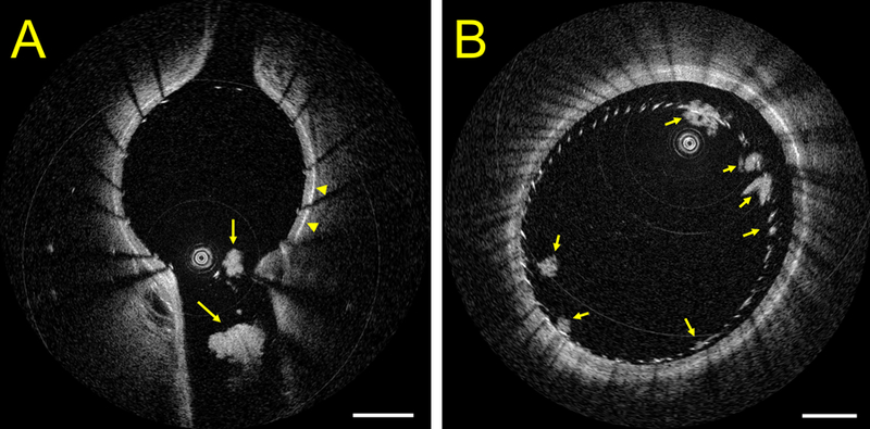
HF-OCT in vivo imaging of neurovascular stenting (Wingspan, Stryker Neurovascular, Fremont CA) in Panel (A), highlighting the presence of multiple thrombi at the level of the ostium of a side-branch in a pig model with no antiplatelet medication. HF-OCT measurements of the longest thrombus diameter are 0.45 mm and 1.05 mm, respectively. The arrowheads point to the vessel external elastic membrane, illustrating the thickness of the vessel wall. Panel B shows a cross-sectional image of a flow-diverter in the same animal model. A flow-diverter malapposition is visible between 11 and 4 o’clock and HF-OCT imaging reveals the presence of multiple clot accumulations on the surface of the device at different locations (arrows). The image scale bars correspond to 1 mm.

HF-OCT in vivo imaging of a coiled aneurysm, side-by-side with x-ray imaging, is shown in Panel (A). HF-OCT cross sectional imaging shows accumulation of thrombus on the surface of the coil wires (arrowheads). The three-dimensional HF-OCT rendering on the right shows additional morphological details of the coil wires that are protruding into the parent artery lumen. A close examination reveals that one of the two wires is floating inside the artery lumen, while the second wire is sitting against the wall of the vessel (arrow). Panel (B) shows a second example of HF-OCT in vivo imaging of a coiled aneurysm. HF-OCT cross-sectional imaging depicts a significant amount of intraluminal thrombus protruding into the parent artery lumen that is not visible on DSA. Panel (C) shows an example of a stented common carotid artery (Precise Pro, Cordis), having a lumen diameter of approximately 5.5 mm. Cross-sectional images scale bars correspond to 1 mm. Three-dimensional visualization is obtained by the means of Osirix MD software (version 9.5.2, Pixmeo Sarl 2016). Limited user processing of the HF-OCT data was applied prior to volumetric rendering.
References
-
- Izatt JA, Hee MR, Swanson EA, Lin CP, Huang D, Schuman JS, et al. Micrometer-scale resolution imaging of the anterior eye in vivo with optical coherence tomography. Arch Ophthalmol 1994;112:1584–1589 - PubMed
-
- Tearney GJ, Brezinski ME, Bouma BE, Boppart SA, Pitris C, Southern JF, et al. In vivo endoscopic optical biopsy with optical coherence tomography. Science 1997;276:2037–2039 - PubMed
Publication types
Grants and funding
LinkOut - more resources
Full Text Sources
