. 2018 Nov 18:2018:4125297.
doi: 10.1155/2018/4125297. eCollection 2018.

Sulforaphane Modulates AQP8-Linked Redox Signalling in Leukemia Cells

Affiliations

Sulforaphane Modulates AQP8-Linked Redox Signalling in Leukemia Cells

Cecilia Prata et al. Oxid Med Cell Longev. .

Abstract

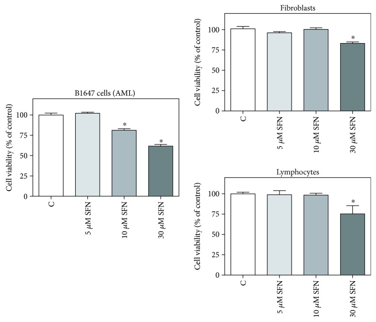
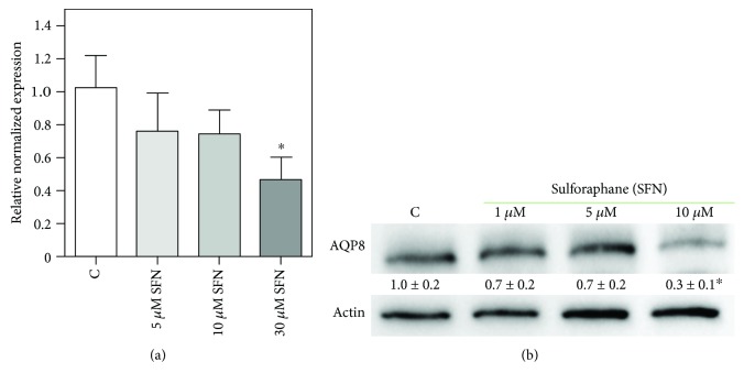
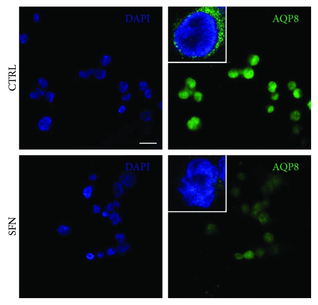
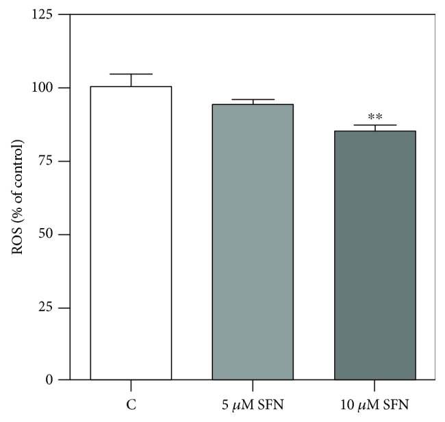
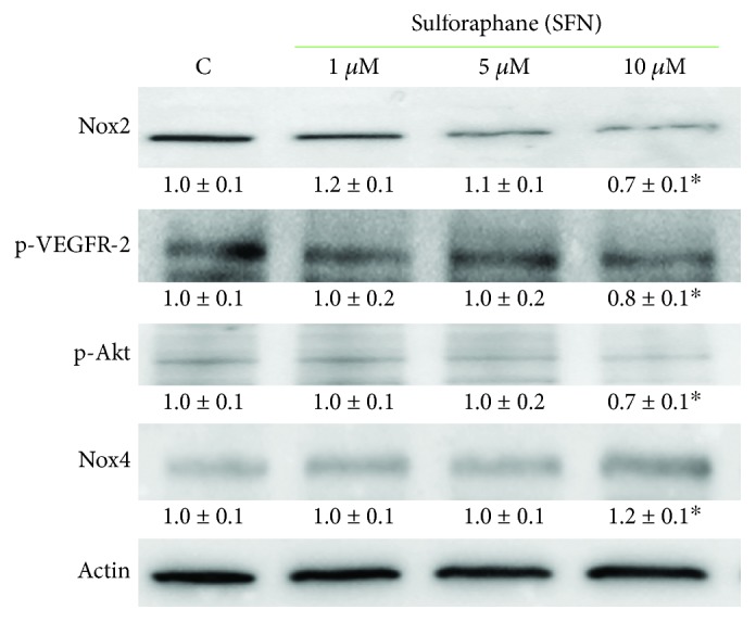
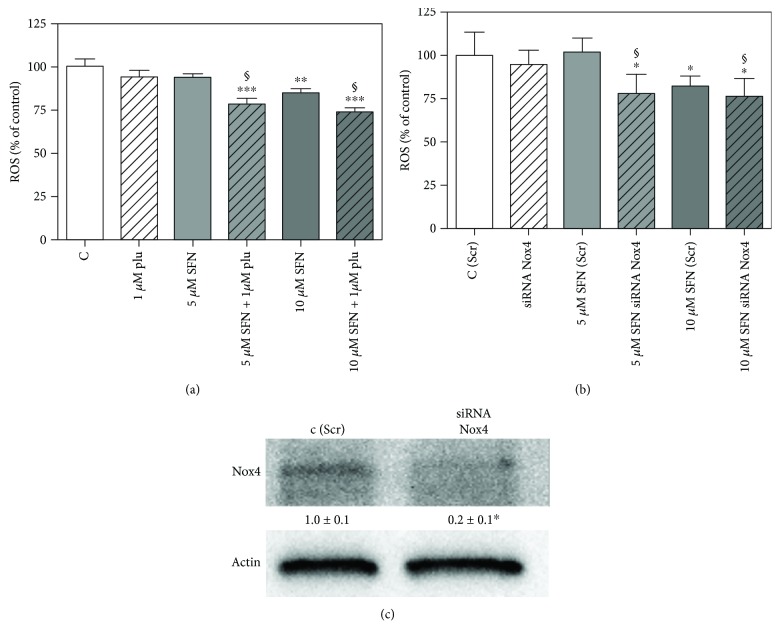
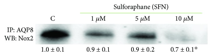
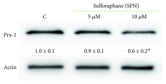
Sulforaphane, a biologically active isothiocyanate compound extracted from cruciferous vegetables, has been shown to exert cytotoxic effects on many human cancer cells, including leukemia. However, the exact molecular mechanisms behind the action of sulforaphane in hematological malignancies are still unclear. Like other cancer cells, leukemia cells produce high level of reactive oxygen species; in particular, hydrogen peroxide derived from Nox family is involved in various redox signal transduction pathways, promoting cell proliferation and survival. Recent evidence show that many tumour cell types express elevated level of aquaporin isoforms, and we previously demonstrated that aquaporin-8 acts as H2O2 transport facilitator across the plasma membrane of B1647 cells, a model of acute myeloid human leukemia. Thus, the control of AQP8-mediated H2O2 transport could be a novel strategy to regulate cell signalling and survival. To this purpose, we evaluated whether sulforaphane could somehow affect aquaporin-8-mediated H2O2 transport and/or Nox-mediated H2O2 production in B1647 cell line. Results indicated that sulforaphane inhibited both aquaporin-8 and Nox2 expression, thus decreasing B1647 cells viability. Moreover, the data obtained by coimmunoprecipitation technique demonstrated that these two proteins are linked to each other; thus, sulforaphane has an important role in modulating the downstream events triggered by the axis Nox2-aquaporin-8. Cell treatment with sulforaphane also reduced the expression of peroxiredoxin-1, which is increased in almost all acute myeloid leukemia subtypes. Interestingly, sulforaphane concentrations able to trigger these effects are achievable by dietary intake of cruciferous vegetables, confirming the importance of the beneficial effect of a diet rich in bioactive compounds.


Figures

Figure 1
Figure 1
Effect of SFN on the viability of transformed and nontransformed human cells. B1647 cells, human lymphocytes or fibroblasts were incubated for 24 h with increasing SFN concentrations. Viability was evaluated by MTT test, as reported in Materials and Methods section. Results are expressed as means ± SD of three independent experiments. Statistical analysis was performed by Bonferroni multiple comparison test following one-way ANOVA. p < 0.05, significantly different from control cells.
Figure 2
Figure 2
Effect of SFN on AQP8 expression in B1647 cell line. B1647 cells were incubated for 24 h with different SFN concentrations and (a) RNA was extracted from the cells and samples subjected to RT-PCR analysis using specific primers as described in Materials and Methods section. Normalized expression levels were calculated relative to control cells according to the 2-∆∆Cq method; (b) proteins were extracted, separated by SDS-PAGE, transferred to nitrocellulose membrane, and immunoassayed using anti-AQP8 and anti-β-actin antibodies as reported in Materials and Methods section. Immunoblot is the representative of three independent experiments, and densitometric analysis, normalized to β-actin, is expressed as fold decrease with respect to control. p < 0.05, significantly different from control cells.
Figure 3
Figure 3
Effect of SFN on AQP8 content in plasma membrane of B1647 cell line. Representative confocal images of B1647 cells treated (SFN) or not (CTRL) with 10 μM sulforaphane for 24 h and labelled with DAPI (blue) and anti-aquaporin 8, AQP8, (green). Cells were not permeabilized in order to exclude intracellular signals. Scale bar = 10 μm. Triple magnification of representative superimposed 3 central slices is shown in white squares. Images were acquired by Nikon A1 confocal laser scanning microscope (Nikon Instruments, Japan). The results are the representative of two independent experiments.
Figure 4
Figure 4
Effect of SFN on intracellular ROS level in B1647 cell line. B1647 cells were incubated for 24 h with different SFN concentrations. Intracellular ROS level was evaluated as DCF fluorescence as reported in Materials and Methods section. Data are expressed as % of control and represent means ± SD of at least three independent experiments. Data were analysed by one-way ANOVA followed by Bonferroni's test. ∗∗p < 0.01, significantly different from control cells.
Figure 5
Figure 5
Effect of SFN on Nox2 and Nox4 expression and phosphorylation level of VEGFR-2 and Akt in B1647 cell line. B1647 cells were incubated for 24 h with different SFN concentrations. At the end of incubation, cells were lysed, and proteins were separated by SDS-PAGE, transferred to nitrocellulose membrane, and immunoassayed using specific antibodies as reported in Materials and Methods section. Immunoblots are the representative of three independent experiments, and densitometric analysis, normalized to β-actin, is expressed as fold decrease with respect to control. p < 0.05, significantly different from control cells.
Figure 6
Figure 6
Effect of SFN on intracellular ROS level in B1647 cell line after Nox4 inhibition or silencing. (a) B1647 cells were incubated for 24 h with different SFN concentrations, then treated or not with 1 μM plumbagin for 30 min. (b) B1647 cells were transfected with specific siRNA against Nox4 or a random RNA sequence (scrambled) as negative control, C (Scr). 24 h after transfection with siRNA, B1647 cells were incubated for 24 h with different SFN concentrations. Intracellular ROS level was then evaluated as DCF fluorescence as reported in Materials and Methods section. Data are expressed as % of control and represent means ± SD of three independent experiments. Data were analysed by one-way ANOVA followed by Bonferroni's test. ∗∗∗p < 0.001; ∗∗p < 0.01; p < 0.05, significantly different from relative control cells. §p < 0.05, significantly different from the corresponding bars in the absence of plumbagin (a) or in Nox4-silenced cells (b). (c) B1647 cells were transfected by electroporation with siRNA against Nox4 or a random RNA sequence (scrambled) as negative control. Effect of RNA interference of Nox4 was confirmed by Western blot analysis with specific antibodies against Nox4.
Figure 7
Figure 7
Effect of SFN on the interaction between AQP8 and Nox2 in B1647 cell line. B1647 cells were incubated for 24 h with different SFN concentrations. At the end of incubation, cells were subjected to immunoprecipitation with anti-AQP8. Proteins were then extracted, separated by SDS-PAGE, immunoblotted, and revealed for anti-Nox2 as described in Materials and Methods section. Immunoblot is the representative of three independent experiments, and densitometric analysis is expressed as fold decrease with respect to control. p < 0.05, significantly different from control cells.
Figure 8
Figure 8
Effect of SFN on peroxiredoxin-1 (Prx-1) in B1647 cell line. B1647 cells were incubated with 5 or 10 μM SFN for 24 h. At the end of incubation, cells were lysed, and proteins were separated by SDS-PAGE, immunoblotted, and revealed for anti-Prx-1 as reported in Materials and Methods section. Immunoblot is the representative of three independent experiments, and densitometric analysis, normalized to β-actin, is expressed as fold decrease with respect to control. p < 0.05, significantly different from control cells.

References

    1. Singh A. K., Sharma N., Ghosh M., Park Y. H., Jeong D. K. Emerging importance of dietary phytochemicals in fight against cancer: role in targeting cancer stem cells. Critical Reviews in Food Science and Nutrition. 2016;57(16):3449–3463. doi: 10.1080/10408398.2015.1129310. - DOI - PubMed
    1. Lenzi M., Fimognari C., Hrelia P. Sulforaphane as a promising molecule for fighting cancer. Cancer Treatment and Research. 2014;159:207–223. doi: 10.1007/978-3-642-38007-5_12. - DOI - PubMed
    1. Angeloni C., Leoncini E., Malaguti M., Angelini S., Hrelia P., Hrelia S. Modulation of phase II enzymes by sulforaphane: implications for its cardioprotective potential. Journal of Agricultural and Food Chemistry. 2009;57(12):5615–5622. doi: 10.1021/jf900549c. - DOI - PubMed
    1. Tarozzi A., Angeloni C., Malaguti M., Morroni F., Hrelia S., Hrelia P. Sulforaphane as a potential protective phytochemical against neurodegenerative diseases. Oxidative Medicine and Cellular Longevity. 2013;2013:10. doi: 10.1155/2013/415078.415078 - DOI - PMC - PubMed
    1. Briones-Herrera A., Eugenio-Pérez D., Reyes-Ocampo J. G., Rivera-Mancía S., Pedraza-Chaverri J. New highlights on the health-improving effects of sulforaphane. Food & Function. 2018;9(5):2589–2606. doi: 10.1039/C8FO00018B. - DOI - PubMed

LinkOut - more resources