Review
. 2018 Nov 22:17:1-13.
doi: 10.1016/j.csbj.2018.11.004. eCollection 2019.

Fates of CD8+ T cells in Tumor Microenvironment

Affiliations
Review

Fates of CD8+ T cells in Tumor Microenvironment

Nomathamsanqa Resegofetse Maimela et al. Comput Struct Biotechnol J. .

Abstract

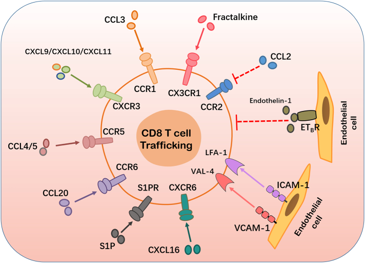
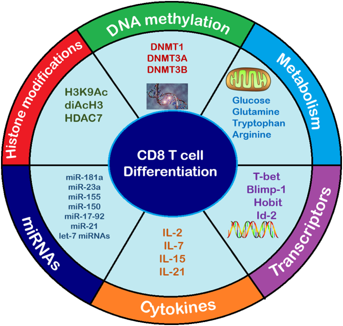
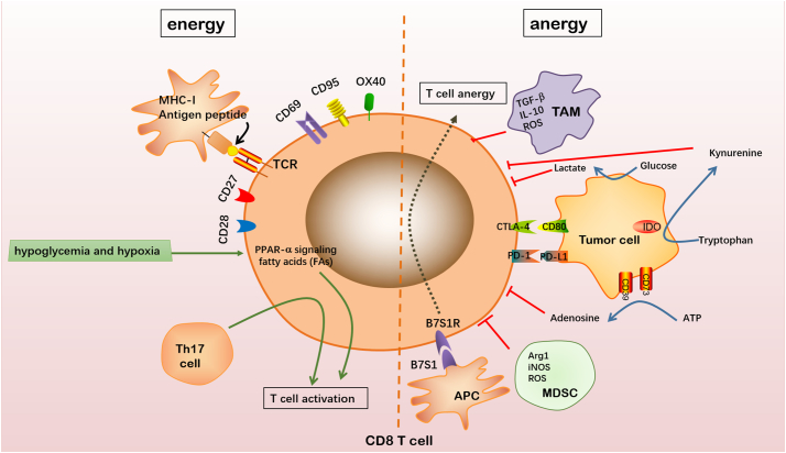
Studies have reported a positive correlation between elevated CD8+ T cells in the tumor microenvironment (TME) and good prognosis in cancer. However, the mechanisms linking T cell tumor-infiltration and tumor rejection are yet to be fully understood. The cells and factors of the TME facilitate tumor development in various ways. CD8+ T cell function is influenced by a number of factors, including CD8+ T cell trafficking and localization into tumor sites; as well as CD8+ T cell growth and differentiation. This review highlights recent literature as well as currently evolving concepts regarding the fates of CD8+ T cells in the TME from three different aspects CD8+ T cell trafficking, differentiation and function. A thorough understanding of factors contributing to the fates of CD8+ T cells will allow researchers to develop new strategies and improve on already existing strategies to facilitate CD8+ T cell mediated anti-tumor function, impede T cell dysfunction and modulate the TME into a less immunosuppressive TME.

Keywords: CD8+ T cell differentiation; CD8+ T cell fates; CD8+ T cell trafficking; T cell exhaustion; Tumor microenvironment.


Figures

Fig. 1
Fig. 1
Factors affecting CD8+ T cell trafficking and localization. CD8+ T cells in the peripheral blood are recruited or trafficked locally into the TME in response to the binding of various secreted chemokines (CCL3, CXCL9, CXCL10, CXCL11, CCL4, CCL5, CCL20, and CXCL16) and their corresponding receptors expressed on tumor cells and stromal cells of the TME. CD8+ T cell trafficking is positively influenced by the interaction between adhesion molecules (ICAM-1, VCAM-1) expressed by endothelial cells and their corresponding receptors which are expressed on the surface of CD8+ T cells. The successful binding of the chemokine ligand fractalkine and CX3CR1 facilitates CD8+ T cell trafficking. The interaction between sphingosine-1-phosphate (S1P) and its corresponding receptors importantly facilitate CD8+ T cell trafficking. The above mentioned receptor-ligand interactions are marked with a solid line and arrow because they promote CD8 T cell trafficking. The binding of endothelin-1 to ETB, its corresponding receptor on endothelial cells, reduces ICAM-1 expression, thus preventing T cell adhesion and subsequently preventing CD8+ T cell trafficking. The chemokine-chemokine receptor interaction between CCL2 and CCR2 inhibits CD8+ T cell trafficking. The later mentioned interactions between endothelin-1 and ETB as well as CCL2 and CCR2 are marked as dotted lines because their interactions inhibit CD8+ T cell trafficking.
Fig. 2
Fig. 2
Factors promoting CD8+ T cell differentiation. CD8+ T cell differentiation and function is promoted by various factors and cellular changes. Metabolic factors such as glucose and amino acids (Glutamine, Tryptophan and Arginine) are used as primary or secondary sources of energy for CD8+ T cell differentiation and function. Transcription factors T-bet, Blimp-1, Hobit and ID2 promote CD8+ T cell differentiation. The presence of cytokines IL-2, IL-7, IL-15 and IL-21 promote CD8+ T cell activation and differentiation. miRNAs such as miR-181a, miR-23a, miR-155, miR-150, miR-17-92, miR-21 and let-7 miRNAs facilitate CD8+ T cell differentiation. Epigenetic changes such as certain histone modifications (H3K9Ac, diAcH3 and HDAC7) and DNA methylations (DNMT1, DNMT3A and DNMT3B) have been identified to facilitate CD8+ T cell differentiation.
Fig. 3
Fig. 3
Factors influencing T cell activation. The formation of the antigenic peptide–MHC-1 complex is a necessary process governing CD8+ T cell activation. Naïve CD8+ T cells may subsequently be activated through their interaction with molecules and cytokines facilitating the activation of their pathways. CD27, CD28, CD69, CD95 and OX40 are costimulatory molecules expressed on CD8+ T cells. The activation of CD8+ T cells is facilitated when any of these costimulatory molecules bind to their respective ligands on APCs. Hypoglycemia and hypoxia lead to increased signaling of peroxisome proliferator-activated receptor (PPAR)-α, facilitating energy generation through fatty acid catabolism and prioritizing CD8+ T cell activation. Th17 cell, a helper T cell, also facilitates CD8+ T cell activation. Naïve CD8+ T cells may be inactivated by the presence of immunosuppressive cells or factors of the TME; interactions with tumor cells; and effects from interactions with other APCs. TAMs secrete TGF-β, IL-10 and ROS to inhibit T cell activation. MDSCs secrete Arg1, iNOS and ROS to inhibit CD8+ T cell activation. Tumor cells expressing PD-L1 or CD80 receptors may bind to corresponding molecules PD-1 and CTLA-4 respectively, causing CD8+ T cell inactivation. Tumor cells may express CD39 and CD73 on their surface facilitating the metabolism of extracellular ATP into AMP and finally into adenosine which inhibits CD8+ T cell activation. Tryptophan is converted into kynurenine through the function of IDO which is produced by the tumor cell. Kynurenine inhibits CD8+ T cell activation. Tumor cells metabolize glucose into lactate and lactate production inhibits CD8+ T cell activation. APCs may express the B7S1 molecule which inhibits CD8+ T cell activation, upon binding to its receptor B7S1R.

References

    1. Nolz J.C. Molecular mechanisms of CD8+ T cell trafficking and localization. Cell Mol Life Sci. 2016;72:2461–2473. - PMC - PubMed
    1. Kim P.S., Ahmed R. Features of responding T cells in cancer and chronic infection. Curr Opin Immunol. 2010;22:223–230. - PMC - PubMed
    1. Kmiecik J. Elevated CD3+ and CD8+ tumor-infiltrating immune cells correlate with prolonged survival in glioblastoma patients despite integrated immunosuppressive mechanisms in the tumor microenvironment and at the systemic level. J Neuroimmunol. 2013;264:71–83. - PubMed
    1. Piersma S.J. High number of intraepithelial CD8+ tumor-infiltrating lymphocytes is associated with the absence of lymph node metastases in patients with large early-stage cervical cancer. Cancer Res. 2007;67:354–361. - PubMed
    1. Bellone M., Calcinotto A. Ways to enhance lymphocyte trafficking into tumors and fitness of tumor infiltrating lymphocytes. Front Oncol. 2013;3:1–15. - PMC - PubMed